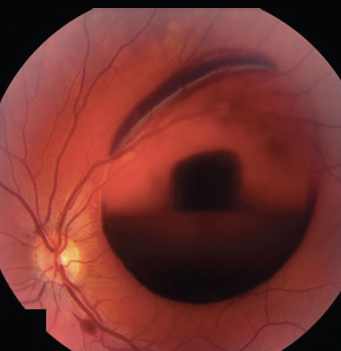
Boat-shaped hemorrhage is a preretinal bleed with a flat upper border and a curved lower edge.
The blood usually lies beneath the posterior hyaloid or the internal limiting membrane (ILM).
Because the sign localizes the blood, it should prompt a focused workup.
What Boat-Shaped Hemorrhage Looks Like
On fundus examination, the lesion forms a dark red pool with a straight, horizontal top.
The lower margin curves like a keel, which explains the name.
Also, the blood hides the retinal vessels beneath it, while the nearby retina may look normal.
Why the Fluid Level Forms
The blood collects in a confined space in front of the retina, where it can settle.
Gravity then pulls red cells downward, while the plasma stays above them.
As a result, an upright patient shows a horizontal interface.
The curved lower edge probably reflects the boundary of the detached hyaloid or ILM.
Fundus Explorer Pro
Photograph the retinal findings described here with the phone already in your pocket — 22 D optics and built-in illumination in one handheld unit.
From Choroida — the team behind this siteSubhyaloid or Sub-ILM Blood
Ophthalmoscopy cannot always separate the two locations.
Sub-ILM blood often forms a taut dome with a glistening surface.
In contrast, subhyaloid blood may shift with posture.
This difference affects surgery, because a sub-ILM collection may need ILM peeling.
Boat-Shaped Hemorrhage and Other Bleeds
Flame hemorrhages follow the nerve fiber layer, while dot and blot hemorrhages sit deeper.
Subretinal blood lies beneath the retinal vessels and often looks darker.
Vitreous hemorrhage, however, blurs the fundus view more diffusely (see vitreous hemorrhage).
Common Causes of Boat-Shaped Hemorrhage
First, let the history guide the search:
- Valsalva retinopathy after straining or vomiting
- Proliferative diabetic retinopathy
- Retinal vein occlusion with neovascularization
- Retinal arterial macroaneurysm
- Terson syndrome after intracranial hemorrhage
- Leukemia, severe anemia or another blood disorder
- Blunt trauma or abusive head trauma in children
History and Examination
First, ask what the patient was doing when the vision changed.
Next, record diabetes, hypertension, anticoagulant use, bruising and recent trauma.
Patients with macular involvement report sudden, painless central vision loss.
Then dilate both eyes, because the fellow eye often provides the clue.
For example, new vessels or laser scars point toward proliferative diabetic retinopathy.
OCT in Boat-Shaped Hemorrhage
OCT shows reflective material in front of the retina, with shadowing beneath it.
However, dense blood can hide the fovea.
Therefore, repeat the scan as the blood clears.
When blood blocks the view, B-scan ultrasound also helps exclude retinal detachment.
Boat-Shaped Hemorrhage as a Red Flag
Severe headache, confusion or reduced consciousness requires urgent neurological assessment.
Terson syndrome may be the first ocular clue to subarachnoid hemorrhage.
Similarly, pallor, fever or unexplained bruising calls for prompt hematological testing.
Never attribute the bleed to Valsalva until the history supports it.
Observing a Boat-Shaped Hemorrhage
Observation suits small bleeds that spare the fovea.
Sleeping with the head elevated may also help blood settle away from the macula.
However, reabsorption can take weeks or months.
During follow-up, check vision, bleed size and any new neovascularization.
Nd:YAG Hyaloidotomy
A laser opening in the posterior wall lets trapped blood drain into the vitreous.
Case series report rapid visual recovery, although randomized evidence is lacking.
This option works best for a fresh, sizable premacular pool (see Nd:YAG hyaloidotomy).
However, risks include macular hole, retinal detachment and epiretinal membrane.
Vitrectomy for Persistent Blood
Vitrectomy clears dense or nonresolving blood and allows ILM peeling when needed.
It also suits eyes with traction or failed laser drainage.
In Terson syndrome, published series describe useful visual recovery.
However, surgery carries risks such as cataract, retinal tears and endophthalmitis.
Treating the Underlying Cause
Drainage clears the visual axis, but it leaves the bleeding source untreated.
Therefore, proliferative disease still needs panretinal photocoagulation or anti-VEGF therapy.
Anti-VEGF injections may also carry traction risks when fibrovascular tissue is present.
Similarly, a blood disorder needs hematology care.
Counseling Patients
Explain that vision can improve as the blood clears.
However, the final outcome depends on the cause and the underlying macula.
Also, advise the patient to report increasing floaters, flashes or a curtain.
Follow-Up After Boat-Shaped Hemorrhage
First, examine the patient again within one to two weeks, because the bleed may enlarge or rupture into the vitreous.
Next, document the size of the pool with serial photographs or OCT.
Then look for residual epiretinal membrane or macular changes once the blood clears.
Finally, schedule systemic review when the cause remains unclear.
Pitfalls to Avoid
- Do not label every bleed as Valsalva without a compatible history
- Avoid laser when the blood has clotted
- Never skip the fellow eye examination
- Recheck the macula once the blood clears


Document what you see
Two smartphone imaging tools built for everyday clinic use — one for the slit lamp, one for the fundus.
From Choroida — the team behind this siteReferences
- Duane TD. Valsalva hemorrhagic retinopathy. Trans Am Ophthalmol Soc. 1972;70:298-313.
- Ulbig MW, Mangouritsas G, Rothbauer HH, et al. Long-term results after drainage of premacular subhyaloid hemorrhage into the vitreous with a pulsed Nd:YAG laser. Arch Ophthalmol. 1998;116:1465-1469.
- Durukan AH, Kerimoglu H, Erdurman C, et al. Long-term results of Nd:YAG laser treatment for premacular subhyaloid haemorrhage owing to Valsalva retinopathy. Eye. 2008;22:214-218.
- Kuhn F, Morris R, Witherspoon CD, Mester V. Terson syndrome: results of vitrectomy and the significance of vitreous hemorrhage in patients with subarachnoid hemorrhage. Ophthalmology. 1998;105:472-477.
Test yourself
A few questions straight from this article.
-
A boat-shaped hemorrhage usually lies in which location?
Boat-shaped hemorrhage is a preretinal bleed that usually lies beneath the posterior hyaloid or the internal limiting membrane, so the sign localizes the blood. -
Why does a boat-shaped preretinal hemorrhage have a flat, horizontal upper border?
Blood in a confined preretinal space settles as gravity pulls red cells down while plasma stays above, giving a horizontal interface in an upright patient. -
Which feature suggests a sub-ILM rather than subhyaloid location for a boat-shaped hemorrhage?
Sub-ILM blood often forms a taut, glistening dome, while subhyaloid blood may shift with posture; a sub-ILM collection may need ILM peeling. -
What distinguishes subretinal blood from a boat-shaped preretinal hemorrhage?
Subretinal blood lies beneath the retinal vessels and often looks darker, whereas a preretinal boat-shaped bleed hides the vessels beneath it. -
A patient with a boat-shaped hemorrhage has severe headache and confusion. Which diagnosis must be considered?
Terson syndrome may be the first ocular clue to subarachnoid hemorrhage, so severe headache, confusion or reduced consciousness needs urgent neurological assessment. -
Which boat-shaped hemorrhage is most suited to observation?
Observation suits small bleeds that spare the fovea, though reabsorption can take weeks or months and follow-up should check for new neovascularization. -
Nd:YAG hyaloidotomy works best for which type of boat-shaped hemorrhage?
A laser opening in the posterior wall lets trapped blood drain into the vitreous, and it works best for a fresh, sizable premacular pool. -
Which complication is a recognized risk of Nd:YAG hyaloidotomy for premacular hemorrhage?
Risks of Nd:YAG hyaloidotomy include macular hole, retinal detachment and epiretinal membrane. -
After a boat-shaped hemorrhage from proliferative diabetic retinopathy is drained, what further treatment is still needed?
Drainage clears the visual axis but leaves the bleeding source untreated, so proliferative disease still needs panretinal photocoagulation or anti-VEGF therapy. -
Why should a patient with a boat-shaped hemorrhage be re-examined within one to two weeks?
Early review within one to two weeks is advised because the bleed may enlarge or break into the vitreous; its size is then followed with photographs or OCT.

Writes and reviews the clinical articles on Educate. Every post is checked against peer-reviewed sources before it goes live.