Vitreous snowballs are discrete, yellow-white aggregates of inflammatory cells suspended in the vitreous.
They usually settle in the inferior vitreous cavity, close to the retinal periphery.
Therefore, they point the examiner toward intermediate or posterior segment inflammation.
What Vitreous Snowballs Are
Histologically, snowballs contain epithelioid cells, lymphocytes and sometimes multinucleated giant cells.
As a result, they behave like small granulomas floating in the gel.
Gravity then pulls them downward, so they cluster inferiorly.
In some eyes they line up in a chain, often called a string of pearls.
Snowbanks Versus Vitreous Snowballs
Snowbanks are white exudative plaques over the inferior pars plana and peripheral retina.
They consist of fibroglial tissue, inflammatory cells and new vessels.
Snowballs float, while snowbanks stay fixed to the vitreous base.
Also, snowbanks usually signal a more chronic and often more severe course.
Fundus Explorer Pro
Photograph the retinal findings described here with the phone already in your pocket — 22 D optics and built-in illumination in one handheld unit.
From Choroida — the team behind this siteHow Patients Present
Most patients report floaters and mild blurring, often in both eyes.
However, vision may fall sharply once cystoid macular edema develops.
Examining for Vitreous Snowballs
Start at the slit lamp and grade anterior vitreous cells and haze.
Next, use indirect ophthalmoscopy with scleral depression to view the inferior periphery.
Snowbanks sit far anteriorly, so a posterior pole view often misses them.
Finally, look for periphlebitis, macular edema and disc swelling.
Grading and Classification
The SUN Working Group defines intermediate uveitis by the vitreous as the primary site of inflammation.
In addition, it reserves the term pars planitis for cases with snowbanks or snowballs.
That label also requires the absence of infection or systemic disease.
Therefore, snowballs alone never complete the diagnosis.
Pars Planitis and Vitreous Snowballs
Pars planitis is the classic idiopathic cause of snowballs and snowbanks.
It is often bilateral and asymmetric, with a chronic relapsing course.
Complications include macular edema, epiretinal membrane, cataract and peripheral neovascularization (see pars planitis in young patients).
Sarcoidosis
Sarcoid uveitis classically produces snowballs and string of pearls opacities in the inferior vitreous.
Also look for mutton fat keratic precipitates, iris nodules and tent shaped peripheral anterior synechiae.
Periphlebitis with candle wax drippings strongly supports the diagnosis (see candle wax drippings in sarcoid).
The IWOS criteria then combine these signs with imaging and biopsy results.
Candida and Infectious Vitreous Snowballs
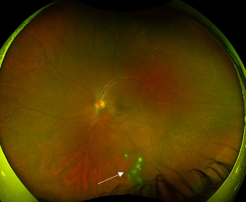
Candida endophthalmitis produces fluffy white vitreous balls, sometimes in a string of pearls pattern.
Usually, a white chorioretinal lesion sits nearby and seeds the vitreous.
Risk factors include intravenous drug use, indwelling catheters, parenteral nutrition, recent surgery and immunosuppression.
Because steroids worsen fungal infection, exclude candida before any steroid injection.
Other Causes in the Differential
- Multiple sclerosis associated intermediate uveitis
- Syphilis, which can mimic almost any uveitis pattern
- Tuberculosis, especially in endemic regions
- Lyme disease in exposed patients
- Toxocariasis with a peripheral granuloma in children
- Vitreoretinal lymphoma in older adults
Masquerade Behind Vitreous Snowballs
Vitreoretinal lymphoma usually affects patients over 50 years.
Its cells form sheets and clumps that can resemble snowballs.
However, the response to steroids stays partial or transient.
Subretinal infiltrates on OCT should then prompt diagnostic vitrectomy.
Workup for Vitreous Snowballs
Tailor tests to the history and exam, then add a short baseline panel.
- Treponemal syphilis serology
- Interferon gamma release assay for tuberculosis
- Serum ACE and lysozyme
- Chest radiograph or chest CT for hilar adenopathy
- Blood cultures and fungal workup when candida is suspected
Imaging
OCT detects cystoid macular edema and epiretinal membrane early.
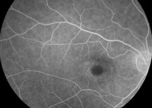
Fluorescein angiography shows peripheral vascular leakage, disc leakage and petaloid macular edema.
Widefield angiography also reveals peripheral leakage that the exam may miss.
Treatment of Inflammatory Disease
Treat the cause first, then the inflammation.
For noninfectious disease, periocular or intravitreal steroids control macular edema in many eyes.
However, steroids raise intraocular pressure and accelerate cataract.
Systemic immunomodulators or adalimumab suit bilateral, recurrent or steroid dependent disease.
Finally, pars plana vitrectomy and peripheral laser help refractory cases and neovascularization.
Treating Candida
Systemic antifungal therapy treats both the eye and the bloodstream infection.
In addition, dense vitritis or macular threat often needs intravitreal antifungal injection.
Vitrectomy also removes organisms and yields material for culture.
Coordinate care with infectious disease specialists, following IDSA guidance.
Counseling Patients
Explain that snowballs are a sign, and the cause still needs testing.
Most idiopathic cases follow a long course with flares and quiet periods.
Also, macular edema drives most vision loss in these eyes.
Therefore, regular follow-up matters even when the eye feels normal.
Pitfalls to Avoid
- Do not inject steroids before excluding candida, syphilis and tuberculosis
- Never skip scleral depression when snowballs are present
- Avoid labeling pars planitis without a negative workup
- Suspect lymphoma when an older patient responds poorly to steroids
For the latest studies, see the PubMed results for vitreous snowballs.


Document what you see
Two smartphone imaging tools built for everyday clinic use — one for the slit lamp, one for the fundus.
From Choroida — the team behind this siteReferences
- Jabs DA, Nussenblatt RB, Rosenbaum JT; SUN Working Group. Standardization of uveitis nomenclature for reporting clinical data. Am J Ophthalmol. 2005;140:509-516.
- SUN Working Group. Classification criteria for pars planitis. Am J Ophthalmol. 2021;228:268-274.
- Herbort CP, Rao NA, Mochizuki M. International criteria for the diagnosis of ocular sarcoidosis. Ocul Immunol Inflamm. 2009;17:160-169.
- Pappas PG, Kauffman CA, Andes DR, et al. Clinical practice guideline for the management of candidiasis: 2016 update. Clin Infect Dis. 2016;62:e1-e50.
- Babu BM, Rathinam SR. Intermediate uveitis. Indian J Ophthalmol. 2010;58:21-27.
Test yourself
A few questions straight from this article.
-
What do vitreous snowballs contain histologically?
Snowballs contain epithelioid cells, lymphocytes and sometimes multinucleated giant cells, so they behave like small granulomas floating in the gel. -
How do vitreous snowballs differ physically from snowbanks?
Snowbanks are white exudative plaques over the inferior pars plana and peripheral retina, fixed to the vitreous base, while snowballs float in the gel. -
Where in the eye do vitreous snowballs usually settle?
Gravity pulls snowballs downward, so they usually settle in the inferior vitreous cavity close to the retinal periphery. -
Which examination technique is needed to view the inferior periphery when vitreous snowballs are present?
Indirect ophthalmoscopy with scleral depression views the inferior periphery; snowbanks sit far anteriorly, so a posterior pole view often misses them. -
Besides snowbanks or snowballs, what does the SUN Working Group require for the label pars planitis?
SUN reserves pars planitis for cases with snowbanks or snowballs and no infection or systemic disease, so snowballs alone never complete the diagnosis. -
In Candida endophthalmitis with vitreous snowballs, what usually lies nearby and seeds the vitreous?
Candida produces fluffy white vitreous balls, usually with a nearby white chorioretinal lesion that seeds the vitreous. -
An older adult has snowball-like vitreous clumps with only a partial steroid response. Which diagnosis should be suspected?
Vitreoretinal lymphoma usually affects patients over 50, its cells can resemble snowballs, and the steroid response stays partial or transient, so subretinal infiltrates on OCT should prompt diagnostic vitrectomy. -
In eyes with idiopathic vitreous snowballs, what drives most vision loss?
Macular edema drives most vision loss in these eyes, so regular follow-up matters even when the eye feels normal. -
Which systemic agent suits bilateral, recurrent or steroid-dependent noninfectious disease with vitreous snowballs?
Systemic immunomodulators or adalimumab suit bilateral, recurrent or steroid-dependent disease, while periocular or intravitreal steroids control macular edema in many eyes. -
Which treatment addresses both the eye and the bloodstream in Candida endophthalmitis with vitreous snowballs?
Systemic antifungal therapy treats both the eye and the bloodstream infection; dense vitritis or macular threat often also needs an intravitreal antifungal.

Writes and reviews the clinical articles on Educate. Every post is checked against peer-reviewed sources before it goes live.