Hyphema grading describes how much blood fills the anterior chamber after trauma.
The grade guides admission, follow-up intervals and surgical thresholds.
It also helps clinicians explain risk to patients in plain terms.
How Hyphema Grading Works
Most clinicians use a height-based scale at the slit lamp.
First, sit the patient upright so the blood settles inferiorly.
Then, measure the level in millimeters with the slit beam.
A photo in the chart also makes later comparison easier.
The Hyphema Grading Scale at a Glance
- Microhyphema: suspended red cells without a layered level
- Grade I: blood filling less than one third of the chamber
- Grade II: blood filling one third to one half
- Subtotal (Grade III): more than half, but short of total
- Total (Grade IV): called eight-ball when the clot turns dark
All-fit Slit-Lamp Adapter
Record and share exactly what you see at the slit lamp. One adapter fits any slit lamp or surgical microscope — and any smartphone.
From Choroida — the team behind this siteMicrohyphema in Hyphema Grading
Microhyphema shows only circulating red cells in the aqueous beam.
Therefore, a penlight examination can easily miss it.
However, microhyphema can still rebleed or raise pressure, especially in sickle cell patients.
Layered Hyphema: Grades I to III
Small layered hyphemas usually clear within several days.
As the level rises, blood covers more outflow surface and hides the pupil.
Therefore, IOP elevation becomes more frequent and more sustained.
Once an open globe is excluded, gentle B-scan ultrasound assesses the posterior segment.
Why the Eight-Ball Hyphema Differs
An eight-ball hyphema is a total clot that looks black or purple.
The dark color reflects deoxygenated blood and stagnant aqueous.
Because aqueous barely circulates, the clot clears slowly.
As a result, pressure spikes and corneal staining become likely.
Hyphema Grading and Rebleed Risk
Secondary hemorrhage usually occurs within the first several days, often days 2 to 5.
During this window the clot retracts and lyses.
Larger initial grades generally carry a higher rebleed risk.
Also, a rebleed is often larger than the first bleed.
Published rates vary widely between series, therefore avoid quoting one figure.
Hyphema Grading and IOP
Any grade can raise pressure, although risk climbs with blood volume.
Red cells, clot and debris obstruct the trabecular meshwork.
Then, a large clot may add pupillary block.
Because corneal edema distorts readings, interpret tonometry carefully (see IOP measurement pitfalls).
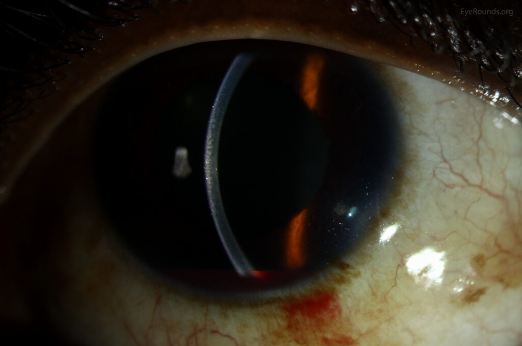
Corneal Blood Staining and Hyphema Grading
Staining begins as a straw-colored haze in the posterior stroma.
It usually follows total hyphema with high IOP or endothelial damage.
Afterward, it clears slowly, starting from the periphery.
In children, central staining can cause amblyopia, so early detection matters.
Sickle Cell Status Changes Every Grade
Sickled red cells pass poorly through the trabecular meshwork.
Therefore, even a microhyphema can cause high IOP in sickle cell disease or trait.
The optic nerve also tolerates pressure poorly in these patients.
For this reason, screen patients of at-risk ancestry early.
Surgical Thresholds by Hyphema Grading
Read and Goldberg advised evacuation for IOP of 50 mmHg for 5 days or 35 mmHg for 7 days.
They also advised surgery for early corneal staining or total hyphema with IOP of 25 mmHg for 6 days.
In sickle cell patients, surgeons often intervene when IOP stays at 24 mmHg or more for over 24 hours.
However, these older thresholds still need individual judgment.
Medical Management Across Grades
Standard care includes a shield, head elevation and reduced activity.
Topical steroids reduce inflammation, while cycloplegics improve comfort.
Avoid aspirin and nonsteroidal anti-inflammatory drugs because they may promote rebleeding.
A Cochrane review found no clear benefit of antifibrinolytics on final visual acuity.
In addition, systemic carbonic anhydrase inhibitors need caution in sickle cell patients.
Follow-Up Intervals by Hyphema Grading
Reliable patients with microhyphema or Grade I hyphema and normal IOP often stay outpatients.
However, higher grades, sickle status or anticoagulation justify daily checks through the rebleed window.
At each visit, regrade the blood, measure IOP and inspect the cornea.
Also, tell patients to report sudden pain, nausea or worse vision at once.
Late Complications After Hyphema
Angle recession can cause glaucoma years later, so perform gonioscopy once the eye settles.
Therefore, lifelong IOP checks are reasonable, as in a glaucoma suspect evaluation.
For a broader review of causes, see the hyphema systematic review.
Pitfalls to Avoid
- Grading a supine patient, which spreads blood and distorts the level
- Missing microhyphema on penlight examination
- Forgetting hemoglobinopathy screening in at-risk patients
- Assuming a low grade means low IOP risk
- Using indentation gonioscopy during the rebleed window


Document what you see
Two smartphone imaging tools built for everyday clinic use — one for the slit lamp, one for the fundus.
From Choroida — the team behind this siteReferences
- Edwards WC, Layden WE. Traumatic hyphema: a report of 184 consecutive cases. Am J Ophthalmol. 1973;75:110-116.
- Read J, Goldberg MF. Comparison of medical treatment for traumatic hyphema. Trans Am Acad Ophthalmol Otolaryngol. 1974;78:OP799-OP815.
- Deutsch TA, Weinreb RN, Goldberg MF. Indications for surgical management of hyphema in patients with sickle cell trait. Arch Ophthalmol. 1984;102:566-569.
- Walton W, Von Hagen S, Grigorian R, Zarbin M. Management of traumatic hyphema. Surv Ophthalmol. 2002;47:297-334.
- Gharaibeh A, Savage HI, Scherer RW, Goldberg MF, Lindsley K. Medical interventions for traumatic hyphema. Cochrane Database Syst Rev. 2019;1:CD005431.
Test yourself
A few questions straight from this article.
-
What defines a microhyphema in hyphema grading?
Microhyphema shows only circulating red cells in the aqueous beam, without a layered level, so a penlight examination can easily miss it. -
Why should a patient sit upright before a hyphema is graded?
Sitting upright lets the blood settle inferiorly, whereas grading a supine patient spreads the blood and distorts the level. -
Which amount of blood in the anterior chamber corresponds to a Grade II hyphema?
Grade I fills less than one third, Grade II one third to one half, subtotal Grade III more than half, and total Grade IV the whole chamber. -
Why does an eight-ball hyphema clear slowly?
An eight-ball hyphema is a total black or purple clot; because aqueous barely circulates it clears slowly, making pressure spikes and corneal staining likely. -
When does secondary hemorrhage after traumatic hyphema usually occur?
Rebleeding usually occurs within the first several days, often days 2 to 5, while the clot retracts and lyses. -
How does hyphema raise intraocular pressure at any grade?
Red cells, clot and debris obstruct the trabecular meshwork, and a large clot may add pupillary block, so risk climbs with blood volume. -
How does corneal blood staining from hyphema first appear?
Staining begins as a straw-colored haze in the posterior stroma, usually after total hyphema with high pressure or endothelial damage, and clears slowly from the periphery. -
Which pressure threshold did Read and Goldberg advise for evacuating a hyphema?
Read and Goldberg advised evacuation for 50 mmHg for 5 days or 35 mmHg for 7 days, although these older thresholds still need individual judgment. -
Which drugs should be avoided in traumatic hyphema because they may promote rebleeding?
Standard care is a shield, head elevation and reduced activity, with aspirin and nonsteroidal anti-inflammatory drugs avoided because they may promote rebleeding. -
Which late complication of hyphema makes gonioscopy worthwhile once the eye settles?
Angle recession can cause glaucoma years later, so gonioscopy is performed once the eye settles and lifelong pressure checks are reasonable.

Writes and reviews the clinical articles on Educate. Every post is checked against peer-reviewed sources before it goes live.