Müller cells are the principal glial cells of the retina.
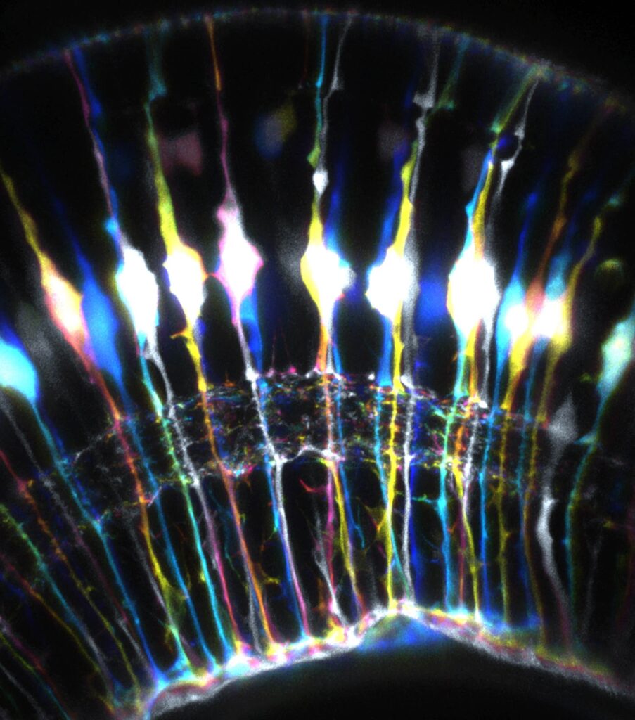
Each cell spans the full thickness from the inner to the outer limiting membrane, so it contacts every neuron and capillary and shapes retinal function at every level.
Anatomy of Müller Cells
The cell body sits in the inner nuclear layer, while its endfeet form the inner limiting membrane and its apical processes form the outer limiting membrane with photoreceptors. Müller cells also account for most of the retinal glial volume, and as a result they act as a scaffold for the whole tissue.
Potassium Buffering
Neural activity releases potassium into the extracellular space, and Müller cells absorb the excess through Kir4.1 channels and release it into the vitreous and blood. This siphoning also keeps the neurons excitable, so failure of these channels disturbs the electroretinogram.
Fundus Explorer Pro
Photograph the retinal findings described here with the phone already in your pocket — 22 D optics and built-in illumination in one handheld unit.
From Choroida — the team behind this siteWater Balance and Aquaporins
Aquaporin 4 channels at the endfeet move water out of the retina with the potassium, and this flow keeps the tissue dry. Loss of the polarity of these channels also leads to swelling, so Müller cells play a role in macular edema.
Glutamate Recycling by Müller Cells
Müller cells take up glutamate from the synapse by transporters, convert it to glutamine with glutamine synthetase, and return it to neurons. This cycle also prevents excitotoxic damage, so impaired uptake raises neuronal injury in ischemia.
Support of the Visual Cycle
Cones can regenerate their visual pigment with the help of Müller cells, and this pathway adds speed to the classic cycle in the pigment epithelium. It also explains how cones adapt in bright light, and Müller cells supply retinoids to cones.
Metabolic Support
Müller cells take up glucose and release lactate for neurons, and they also store glycogen and shield the retina against oxidative stress. They make glutathione as well, and as a result they act as an energy partner to photoreceptors.
Light Guidance
Franze and colleagues showed that Müller cells guide light through the retina like optical fibers, carrying it from the inner surface to the photoreceptors with low loss. This arrangement improves image contrast, and therefore the glial cells also have an optical role.
Barrier Function
Müller cells contribute to the inner blood-retinal barrier together with pericytes and endothelial cells, and they release factors that keep the capillary tight. Injury to the cells also opens the barrier, so gliosis often accompanies leakage.
Reactive Gliosis
In disease, the cells increase glial fibrillary acidic protein and change their shape, and mild gliosis protects neurons while severe gliosis forms scars. The cells can also proliferate on the retinal surface, so gliosis can be a friend or an enemy.
Müller Cells in Macular Holes and Epiretinal Membranes
Gass described a Müller cell cone at the fovea as the center of tissue support, and loss or traction of this cone is linked to hole formation (see full-thickness macular hole). Müller cells also grow along the epiretinal membranes, so they link several vitreoretinal disorders.
Müller Cells in Diabetic Retinopathy
High glucose alters the glutamate cycle and Kir4.1 expression in the glial cells, and early gliosis precedes visible vessel changes. The cells also release VEGF under hypoxia, so they take part in leakage and neovascularization.
Müller Cells in Inherited Disease
Müller cell dysfunction explains the retinal changes in X-linked retinoschisis. Retinoschisin is secreted by photoreceptors and bipolar cells and binds to Müller cells, and mutations disturb the cell contacts and cause splitting of layers, and the negative electroretinogram follows the disruption.
Regeneration
Fish Müller cells dedifferentiate and produce new neurons after injury, but in mammals this ability is very limited. Researchers also try to reactivate it with transcription factors, so the cells are a target for future regenerative therapy.
Clinical Signs Linked to the Cells
OCT shows cystic spaces when Müller cell function fails in edema, and hyperreflective columns can reflect Müller cell processes. The electroretinogram b-wave also depends partly on potassium currents in the cells, so imaging and electrophysiology can reveal glial dysfunction.
Pitfalls to Avoid
- Do not view Müller cells as passive support only
- Never read gliosis as always harmful
- Do not ignore glia when planning internal limiting membrane peeling
- Never assume that animal regeneration data apply directly to humans
Detachment and Gliosis
After detachment, the glial cells proliferate and migrate on the retinal surface, and this process feeds proliferative vitreoretinopathy and epiretinal membranes. Early reattachment also limits gliosis, so speed of repair helps recovery.


Document what you see
Two smartphone imaging tools built for everyday clinic use — one for the slit lamp, one for the fundus.
From Choroida — the team behind this siteReferences
- Bringmann A, Pannicke T, Grosche J, et al. Müller cells in the healthy and diseased retina. Prog Retin Eye Res. 2006;25:397-424.
- Reichenbach A, Bringmann A. New functions of Müller cells. Glia. 2013;61:651-678.
- Franze K, Grosche J, Skatchkov SN, et al. Müller cells are living optical fibers in the vertebrate retina. Proc Natl Acad Sci U S A. 2007;104:8287-8292.
- Gass JD. Müller cell cone, an overlooked part of the anatomy of the fovea centralis: hypotheses concerning its role in the pathogenesis of macular hole and foveomacular retinoschisis. Arch Ophthalmol. 1999;117:821-823.
Test yourself
A few questions straight from this article.
-
In which retinal layer does the cell body of the Müller cell sit?
The Müller cell body sits in the inner nuclear layer. Its endfeet form the inner limiting membrane and its apical processes form the outer limiting membrane, so the cell spans the full retinal thickness. -
Through which channels do retinal Müller cells absorb excess extracellular potassium released by neural activity?
Müller cells absorb excess potassium through Kir4.1 channels and release it into the vitreous and blood. Failure of these channels disturbs the electroretinogram. -
How does loss of aquaporin 4 polarity at Müller cell endfeet contribute to macular edema?
Aquaporin 4 channels at the endfeet move water out of the retina along with potassium, keeping it dry. Loss of their polarity leads to swelling, linking Müller cells to macular edema. -
Which enzyme do retinal Müller cells use to convert synaptic glutamate into glutamine?
Müller cells take up glutamate by transporters, convert it to glutamine with glutamine synthetase and return it to neurons. This prevents excitotoxic damage, so impaired uptake increases neuronal injury in ischemia. -
What optical role did Franze and colleagues demonstrate for retinal Müller cells?
Müller cells carry light from the inner retinal surface to the photoreceptors with low loss, acting like optical fibers. The arrangement improves image contrast. -
Which protein do Müller cells increase during reactive gliosis?
In disease, Müller cells increase glial fibrillary acidic protein and change shape. Mild gliosis protects neurons, whereas severe gliosis forms scars. -
Which foveal structure did Gass describe as the centre of tissue support, whose loss or traction is linked to macular hole formation?
Gass described a Müller cell cone at the fovea as the centre of tissue support. Loss or traction of this cone is linked to hole formation. -
In diabetic retinopathy, how do Müller cells contribute to leakage and neovascularization?
Müller cells release VEGF under hypoxia and so take part in leakage and neovascularization. High glucose also alters their glutamate cycle, and early gliosis precedes visible vessel changes. -
In X-linked retinoschisis, which cells secrete retinoschisin, the protein that binds to Müller cells?
Retinoschisin is secreted by photoreceptors and bipolar cells and binds to Müller cells. Mutations disturb cell contacts, causing splitting of retinal layers and a negative electroretinogram. -
After retinal detachment, which complications does Müller cell proliferation on the retinal surface feed?
After detachment, Müller cells proliferate and migrate on the retinal surface, feeding proliferative vitreoretinopathy and epiretinal membranes. Early reattachment limits this gliosis.

Writes and reviews the clinical articles on Educate. Every post is checked against peer-reviewed sources before it goes live.