
Amniotic membrane transplantation has become one of the more versatile tools in ocular surface reconstruction, used across a genuinely wide range of conditions, and its usefulness across such different clinical scenarios comes down to a consistent set of biological properties this tissue provides regardless of which specific problem it is being applied to.
Why This Tissue Works So Well on the Ocular Surface
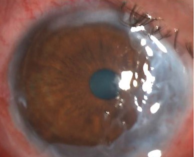
Amniotic membrane, the innermost layer of the placenta, processed and prepared for surgical use, provides a basement membrane that supports epithelial cell migration and adhesion, along with anti-inflammatory, anti-scarring, and anti-angiogenic properties, creating a favorable environment for ocular surface healing across a range of otherwise quite different underlying conditions.
Acute Chemical and Thermal Burns
Applied early in the course of a significant chemical or thermal burn, amniotic membrane can reduce inflammation and scarring during the acute healing phase, and evidence suggests that early application, particularly within the first one to two weeks after a severe burn, can meaningfully reduce the severity of chronic ocular surface sequelae (see Stevens-Johnson syndrome ocular sequelae for a related discussion of this same early-intervention principle applied to a different, though similarly severe, acute ocular surface insult).
All-fit Slit-Lamp Adapter
Record and share exactly what you see at the slit lamp. One adapter fits any slit lamp or surgical microscope — and any smartphone.
From Choroida — the team behind this sitePersistent Epithelial Defects
For corneal epithelial defects that fail to heal with standard supportive measures, amniotic membrane provides a supportive scaffold and anti-inflammatory environment that can promote healing in defects that have proven resistant to more conservative measures alone, offering an intermediate option before considering more invasive surgical approaches.
Limbal Stem Cell Transplantation Support
Amniotic membrane is frequently used as an adjunctive substrate during limbal stem cell transplantation procedures, supporting the transplanted limbal tissue and the migrating epithelial cells during the critical early healing period (see limbal stem cell transplantation techniques for the broader surgical context in which this adjunctive tissue is commonly used).
Pterygium Surgery
Amniotic membrane grafting is an alternative to conjunctival autografting for covering the bare scleral defect after pterygium excision, particularly useful in cases where conjunctival autograft tissue is limited, such as recurrent pterygium surgery or when conjunctival tissue needs to be preserved for potential future glaucoma surgery.
Symblepharon and Conjunctival Reconstruction
Amniotic membrane is used in reconstructing the conjunctival surface and fornices in conditions causing conjunctival scarring and symblepharon formation, including chemical burns and cicatricial conjunctival diseases, helping to restore normal fornix depth and reduce recurrent scarring during the healing process.
Application Techniques
As an Overlay (Patch)
The membrane is placed over the ocular surface, generally secured with sutures, a bandage contact lens, or a specialized ring device, providing a protective, biologically active covering that can be left in place for days to weeks depending on the clinical indication.
As an Inlay (Graft)
The membrane is used to fill or replace a tissue defect directly, providing a structural scaffold integrated more directly into the healing surface, as used in some persistent epithelial defect and reconstructive applications.
Sutureless Application
Some amniotic membrane products are designed for sutureless application, using a specialized ring or adhesive system, offering a faster, simpler application technique particularly useful in an office-based setting for appropriate indications.
Considerations and Limitations
While amniotic membrane transplantation is valuable across these varied applications, it is generally used as part of a broader treatment plan rather than as a standalone cure for the underlying condition, and addressing the primary cause of ocular surface compromise, whatever it may be in a given patient, remains essential alongside the supportive benefits this adjunctive tissue provides.
Practical points on handling the tissue
Amniotic membrane is available as cryopreserved or dehydrated tissue. Cryopreserved membrane retains more of its biological activity but needs frozen storage, while dehydrated membrane can be stored at room temperature and is easier to use in the clinic. The membrane has a basement membrane side and a stromal side, and it is usually placed with the stromal side toward the wound, depending on the technique, so follow the manufacturer's instructions.
Pitfalls
- Do not use the membrane as a substitute for treating infection or underlying disease
- Do not place it over an eye with uncontrolled infection
- Do not forget to monitor the intraocular pressure when steroids are used
- Do not remove sutures too early in an inflamed eye
Record the type of membrane, the batch number, and the position, so that the information is available at follow-up.
Prognosis
Amniotic membrane transplantation, applied appropriately across its various indications, generally supports improved healing, reduced inflammation and scarring, and better overall ocular surface outcomes compared with management without this adjunctive tissue.
Its versatility across such a range of distinct clinical scenarios, acute burns, persistent defects, stem cell transplantation support, pterygium surgery, and conjunctival reconstruction, reflects the genuinely broad utility of its underlying biological properties rather than effectiveness limited to any single specific application.


Document what you see
Two smartphone imaging tools built for everyday clinic use — one for the slit lamp, one for the fundus.
From Choroida — the team behind this siteReferences
- Dua HS, Gomes JA, King AJ, Maharajan VS. The amniotic membrane in ophthalmology. Surv Ophthalmol. 2004;49:51-77.
- Gregory DG. Treatment of acute Stevens-Johnson syndrome and toxic epidermal necrolysis using amniotic membrane: a review of 10 consecutive cases. Ophthalmology. 2011;118:908-914.
- John T. Human amniotic membrane transplantation: past, present, and future. Ophthalmol Clin North Am. 2003;16:43-65.
- Prabhasawat P, Barton K, Burkett G, Tseng SC. Comparison of conjunctival autografts, amniotic membrane grafts, and primary closure for pterygium excision. Ophthalmology. 1997;104:974-985.
Test yourself
A few questions straight from this article.
-
Amniotic membrane used in ocular surface surgery is derived from which structure?
Amniotic membrane is the innermost layer of the placenta, processed and prepared for surgical use on the ocular surface. -
Which property set explains amniotic membrane's usefulness across different ocular surface diseases?
The membrane supplies a basement membrane that supports epithelial migration and adhesion, and carries anti-inflammatory, anti-scarring and anti-angiogenic properties. -
After a severe chemical or thermal burn, early amniotic membrane application is most beneficial within which window?
Evidence suggests application within the first one to two weeks after a severe burn meaningfully reduces the severity of chronic ocular surface sequelae. -
In a corneal epithelial defect resistant to standard supportive measures, what role does amniotic membrane serve?
For defects resistant to conservative measures the membrane gives a supportive scaffold and anti-inflammatory environment, an intermediate step before more invasive surgical approaches. -
How is amniotic membrane used during limbal stem cell transplantation?
It serves as an adjunctive substrate supporting the transplanted limbal tissue and the migrating epithelial cells through the critical early healing period. -
Why may amniotic membrane be chosen over conjunctival autograft after pterygium excision?
Amniotic membrane covers the bare scleral defect when autograft tissue is limited, as in recurrent pterygium or when conjunctiva must be preserved for future glaucoma surgery. -
What is the aim of amniotic membrane grafting in conjunctival scarring with symblepharon?
In conjunctival scarring and symblepharon formation the membrane helps restore normal fornix depth and reduce recurrent scarring during healing. -
How does inlay (graft) amniotic membrane use differ from overlay (patch) use?
As an inlay the membrane fills or replaces a tissue defect and integrates into the healing surface, whereas an overlay sits over the surface as a protective covering. -
What is the main trade-off of cryopreserved versus dehydrated amniotic membrane?
Cryopreserved membrane retains more of its biological activity but requires frozen storage, while dehydrated membrane keeps at room temperature and is easier to use in clinic. -
Where does amniotic membrane transplantation sit within an ocular surface treatment plan?
The membrane is used as part of a broader treatment plan rather than a standalone cure, and addressing the primary cause of ocular surface compromise remains essential.
