
Dacryoadenitis is inflammation of the lacrimal gland, located in the superolateral orbit.
It is easily confused with — but mechanistically and anatomically distinct from — dacryocystitis, which involves the lacrimal sac in the medial canthal region.
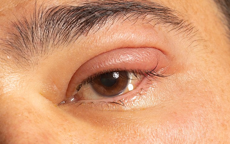
The location of the swelling is the single fastest way to tell the two apart clinically, and getting this distinction right matters directly for the differential diagnosis and workup that follows, since the two conditions point toward almost entirely different sets of underlying causes.
Acute Dacryoadenitis
Acute dacryoadenitis presents with sudden onset of pain, swelling, and erythema over the superolateral orbit, classically producing an “S-shaped” or laterally displaced upper eyelid contour from the swollen gland pushing the lid margin out of its normal curve.
Viral causes (mumps, Epstein-Barr virus, and other common viral pathogens) are more frequent overall, particularly in children, and tend to be self-limited.
Meanwhile, bacterial dacryoadenitis (Staphylococcus and Streptococcus species being typical culprits) is less common but more likely to require targeted antibiotic treatment and, occasionally, drainage if an abscess develops.
Chronic Dacryoadenitis
Chronic dacryoadenitis, generally defined as lacrimal gland inflammation persisting beyond about a month, is far more often related to a systemic inflammatory or infiltrative condition rather than to infection.
Identifying that underlying systemic disease is the central task of the workup once the acute infectious causes have been reasonably excluded.
Because the two forms — acute infectious and chronic systemic — require fundamentally different evaluations and treatments, the duration and character of the swelling (sudden and painful versus gradual and painless) is often the single most useful piece of history for deciding how far to take the initial workup.
All-fit Slit-Lamp Adapter
Record and share exactly what you see at the slit lamp. One adapter fits any slit lamp or surgical microscope — and any smartphone.
From Choroida — the team behind this siteCauses of Chronic Dacryoadenitis
- Sarcoidosis — one of the more classic causes, sometimes bilateral, and worth specific consideration given the broader systemic implications of a new sarcoidosis diagnosis
- IgG4-related disease — an increasingly recognized cause of chronic, often bilateral lacrimal gland enlargement, sometimes as part of a broader multi-organ IgG4-related process
- Sjögren’s syndrome — lacrimal gland involvement as part of the broader autoimmune exocrine gland dysfunction that defines the condition
- Lymphoproliferative disease — orbital lymphoma can present as a slowly enlarging, painless lacrimal gland mass, an important and higher-stakes mimic of benign chronic dacryoadenitis
- Idiopathic orbital inflammation (orbital pseudotumor) specifically involving the lacrimal gland — sometimes called dacryoadenitis-predominant orbital inflammatory syndrome
- Graves’ (thyroid eye) disease, occasionally with lacrimal gland involvement alongside its more classic extraocular muscle findings
Clinical Findings
- Swelling localized to the superolateral orbit/upper eyelid, corresponding to the anatomic location of the lacrimal gland
- An S-shaped ptosis or lid contour deformity in more significant swelling
- Palpable, sometimes tender (in acute disease), enlargement of the palpebral lobe of the lacrimal gland on lid eversion
- Associated conjunctival injection over the gland
- In chronic disease: painless, gradual enlargement, sometimes bilateral, without the acute inflammatory signs of the infectious form
Differential Diagnosis
- Dacryocystitis — swelling centered medially, over the lacrimal sac, rather than superolaterally, discussed in its own dedicated article on this site
- Preseptal or orbital cellulitis — broader periorbital involvement not specifically localized to the lacrimal gland
- Chalazion or hordeolum of the lateral upper lid — more discrete, smaller, and not corresponding specifically to the lacrimal gland’s anatomic location
- Orbital or lacrimal gland tumor (benign pleomorphic adenoma, or malignant lacrimal gland carcinoma) — typically painless and slowly progressive, an important distinction from acute inflammatory dacryoadenitis and one that imaging helps clarify
Diagnostic Evaluation
Acute, classic viral dacryoadenitis in a child with a clear preceding viral illness may not require extensive workup beyond clinical observation.
Chronic, atypical, or recurrent dacryoadenitis warrants orbital imaging (CT or MRI) to characterize the gland and exclude a mass lesion, along with systemic workup tailored to the suspected cause: ACE and chest imaging for sarcoidosis, serum IgG4 levels for IgG4-related disease, and relevant autoantibody testing (SSA/SSB) for Sjögren’s syndrome.
Biopsy is reserved for cases where the diagnosis remains unclear after imaging and serologic workup, or when malignancy is a genuine concern based on the clinical picture, given the added risk and morbidity of a lacrimal gland biopsy compared with less invasive testing.
Bilateral involvement, while not specific to any single cause, is more suggestive of a systemic process (sarcoidosis, IgG4-related disease, Sjögren’s syndrome) than of a localized tumor, and can help prioritize the systemic workup even before results of any individual test are available.
Management
Viral dacryoadenitis is managed supportively, with resolution expected over one to a few weeks as the underlying viral illness resolves.
Bacterial dacryoadenitis is treated with targeted systemic antibiotics, with surgical drainage reserved for cases that develop a frank abscess not responding to antibiotics alone.
Chronic dacryoadenitis is managed by treating the identified underlying systemic condition — corticosteroids and, where needed, steroid-sparing immunosuppression for sarcoidosis or IgG4-related disease, and appropriate oncologic management if a lymphoproliferative process is identified — rather than by treating the lacrimal gland swelling as an isolated local problem.
Long-term follow-up, often shared between ophthalmology and whichever specialist manages the underlying systemic disease, is standard for chronic dacryoadenitis, since gland size and inflammatory activity are useful markers of how well the broader systemic condition is being controlled over time, and a change in the gland can sometimes be the first sign of a flare or relapse in the underlying disease.


Document what you see
Two smartphone imaging tools built for everyday clinic use — one for the slit lamp, one for the fundus.
From Choroida — the team behind this siteReferences
- Bernardini FP, Devoto MH, Croxatto JO. Epidemiology of lacrimal gland tumors. Ophthalmic Plastic and Reconstructive Surgery.
- Andrew NH, Kearney D, Selva D. IgG4-related orbital disease: a meta-analysis and review. Acta Ophthalmologica.
- American Academy of Ophthalmology. Basic and Clinical Science Course, Section 7: Orbit, Eyelids, and Lacrimal System.
