
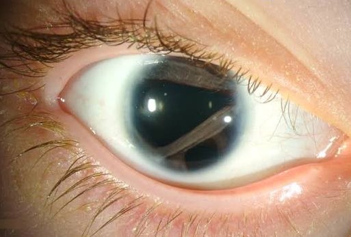
Axenfeld-Rieger syndrome is a spectrum of anterior segment dysgenesis affecting neural crest-derived tissue, producing a combination of corneal, iridic, and angle abnormalities that carries a substantial lifelong glaucoma risk, together with characteristic systemic features, most notably dental and facial anomalies.
The eye findings here are unusually visible and specific, which means a large part of managing this condition well is simply recognising the pattern early enough to start glaucoma surveillance before any damage has occurred, rather than waiting for symptoms that this disease does not reliably produce until late.
Classification Within the Spectrum
- Axenfeld anomaly – posterior embryotoxon with iris strands adherent to the prominent Schwalbe line, without significant iris stromal atrophy
- Rieger anomaly – the ocular findings of Axenfeld anomaly plus iris stromal atrophy, corectopia, and polycoria (multiple pupillary openings)
- Rieger syndrome – Rieger anomaly plus systemic features, most consistently dental hypoplasia or missing teeth, and characteristic midface hypoplasia
Rather than being distinct diseases, these terms describe overlapping points on a single developmental spectrum, and “Axenfeld-Rieger syndrome” is now commonly used as an umbrella term covering the full range, with individual patients falling anywhere along it.
Genetics
Mutations in PITX2 and FOXC1 account for a substantial proportion of identified cases, both genes encoding transcription factors involved in normal anterior segment and craniofacial development, which explains the combination of ocular and dental or facial findings seen in the syndrome.
Inheritance is typically autosomal dominant with variable expressivity, meaning severity can differ considerably even between affected members of the same family, and some cases arise from new mutations without a family history.
All-fit Slit-Lamp Adapter
Record and share exactly what you see at the slit lamp. One adapter fits any slit lamp or surgical microscope — and any smartphone.
From Choroida — the team behind this siteOcular Findings
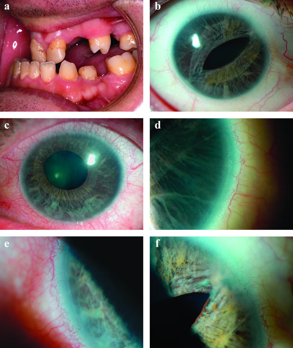
Posterior embryotoxon is essentially universal in this syndrome, visible as a prominent, anteriorly displaced Schwalbe line at the limbus. Iris strands can be seen bridging from the peripheral iris to this line, sometimes distorting the pupil.
Iris stromal thinning, corectopia (a displaced pupil), and polycoria are characteristic of the more severe end of the spectrum, and peripheral anterior synechiae, visible on gonioscopy, reflect the underlying angle dysgenesis that ultimately drives the glaucoma risk in this condition.
Bilateral involvement is typical, though the severity is frequently asymmetric between the two eyes, which is worth remembering when assessing glaucoma risk, since a milder-appearing eye can still carry meaningful risk.
Systemic Findings
- Dental anomalies, including microdontia, hypodontia, and widely spaced or missing teeth, among the most consistent systemic features and often visible simply by asking the patient or family to describe their dental history
- Maxillary hypoplasia and a flattened midface profile
- Redundant periumbilical skin, present in a subset of patients
- Pituitary abnormalities and, rarely, growth hormone deficiency have been reported in some cases, relevant to broader paediatric assessment when the diagnosis is confirmed in a child
Glaucoma Risk
Glaucoma develops in roughly half of patients with Axenfeld-Rieger syndrome, arising from the underlying angle dysgenesis that disrupts normal aqueous outflow, and it can present at any point from infancy through adulthood, which is the central reason lifelong surveillance is needed rather than a fixed period of childhood monitoring.
Because early glaucoma can be asymptomatic, regular intraocular pressure checks and optic disc assessment are essential in every patient with this diagnosis, even those without visible severe iris changes, since disease severity in the iris does not reliably predict glaucoma risk.
Differential Diagnosis
- Isolated posterior embryotoxon, lacking the iris changes, systemic dental findings, and elevated glaucoma risk of the full syndrome
- Peters anomaly, a more severe anterior segment dysgenesis with central corneal opacity rather than the peripheral and iridic findings characteristic of Axenfeld-Rieger syndrome
- Aniridia, distinguished by near-complete absence of the iris rather than the strand adhesions and corectopia seen here
- Congenital ectropion uveae, a different, non-syndromic iris abnormality without the corneal and systemic findings of Axenfeld-Rieger syndrome
Management
Glaucoma Surveillance and Treatment
Regular intraocular pressure monitoring should begin as soon as the diagnosis is suspected and continue for life, since glaucoma can develop at any age. When glaucoma is identified, management follows standard principles for the age of onset, whether that means angle surgery, similar to primary congenital glaucoma, in infancy, or medical and surgical approaches used for adult-onset glaucoma later in life.
Refractive and Amblyopia Management
Refractive error and amblyopia should be assessed and treated using standard paediatric principles, since corneal and angle abnormalities can be associated with astigmatism or anisometropia that, left uncorrected, risks amblyopia independent of the glaucoma itself.
Systemic and Dental Referral
Dental assessment and, where relevant, paediatric evaluation for growth or pituitary concerns should be arranged once the diagnosis is confirmed, since the systemic aspects of this syndrome benefit from coordinated, multidisciplinary care rather than being treated as incidental findings.
Genetic Counselling
Given autosomal dominant inheritance with variable expressivity, genetic counselling is appropriate for affected families, including discussion of the range of severity that can occur even within the same family and screening considerations for at-risk relatives.
Prognosis
Visual outcome depends primarily on how effectively glaucoma is controlled over a patient’s lifetime, since uncontrolled intraocular pressure, not the iris or corneal changes themselves, is what threatens vision in this condition.
With regular surveillance and timely treatment, many patients maintain good vision throughout life, though a meaningful minority develop significant glaucomatous damage, particularly if diagnosis or the start of monitoring is delayed.
Systemic features, particularly dental anomalies, are generally manageable with appropriate dental care and do not independently threaten health, meaning that for most patients, long-term outcome ultimately comes down to how consistently the lifelong glaucoma surveillance this syndrome requires is actually maintained.


Document what you see
Two smartphone imaging tools built for everyday clinic use — one for the slit lamp, one for the fundus.
From Choroida — the team behind this siteReferences
- Shields MB. Axenfeld-Rieger syndrome: a spectrum of developmental disorders. Survey of Ophthalmology. 1985.
- Tumer Z, Bach-Holm D. Axenfeld-Rieger syndrome and spectrum of PITX2 and FOXC1 mutations. European Journal of Human Genetics. 2009.
- Ito YA, Walter MA. Genomics and anterior segment dysgenesis: a review. Clinical and Experimental Ophthalmology. 2014.
- Axenfeld-Rieger Syndrome. EyeWiki, American Academy of Ophthalmology.
- Axenfeld-Rieger Syndrome. StatPearls, NCBI Bookshelf.
Test yourself
A few questions straight from this article.
-
In the Axenfeld-Rieger spectrum, which finding upgrades Axenfeld anomaly to Rieger anomaly?
Rieger anomaly adds iris stromal atrophy, corectopia and polycoria to the Axenfeld findings; systemic features are what then define Rieger syndrome. -
Mutations in which two transcription factor genes account for a substantial proportion of Axenfeld-Rieger syndrome cases?
Both encode transcription factors governing anterior segment and craniofacial development, which explains why ocular findings travel with dental and facial anomalies. -
What inheritance pattern does Axenfeld-Rieger syndrome typically follow?
Severity can differ considerably between affected members of one family, and some cases arise as new mutations with no family history at all. -
Which anterior segment finding is essentially universal in Axenfeld-Rieger syndrome?
It appears as a prominent, anteriorly displaced Schwalbe line, often with iris strands bridging across to it from the peripheral iris. -
Which gonioscopic finding in Axenfeld-Rieger syndrome reflects the angle dysgenesis behind its glaucoma risk?
Peripheral anterior synechiae seen on gonioscopy mark the underlying angle dysgenesis that disrupts aqueous outflow and ultimately drives glaucoma. -
Approximately what proportion of patients with Axenfeld-Rieger syndrome develop glaucoma?
Glaucoma arises in roughly half of patients and can appear at any point from infancy through adulthood, which is why surveillance must be lifelong. -
Which systemic feature is the most consistent extraocular finding in Axenfeld-Rieger syndrome?
Microdontia, hypodontia and widely spaced or missing teeth are among the most consistent systemic features, often uncovered by asking about dental history. -
Which anterior segment dysgenesis is distinguished from Axenfeld-Rieger syndrome by a central corneal opacity?
Peters anomaly is a more severe dysgenesis centred on the cornea, whereas Axenfeld-Rieger findings are peripheral and iridic. -
How should intraocular pressure be monitored in a patient with Axenfeld-Rieger syndrome?
Glaucoma can develop at any age and is often asymptomatic early, and the severity of iris change does not reliably predict who is at risk. -
What primarily determines long-term visual outcome in Axenfeld-Rieger syndrome?
Uncontrolled intraocular pressure, not the iris or corneal changes themselves, is what threatens vision across a patient's lifetime.

Writes and reviews the clinical articles on Educate. Every post is checked against peer-reviewed sources before it goes live.


