
Iris heterochromia is a difference in iris colour, either between the two eyes or within a single iris, and while it is frequently a harmless constitutional trait, it belongs on a differential that also includes Horner syndrome, chronic uveitis, iron deposition, and ocular tumours.
The clinical task is rarely difficult once approached systematically: distinguishing longstanding, stable, isolated heterochromia from newly acquired or changing pigmentation, since that single distinction does most of the work of separating the benign cases from the ones that need a workup.
Types of Heterochromia
- Complete heterochromia iridum – one iris is entirely a different colour from the other
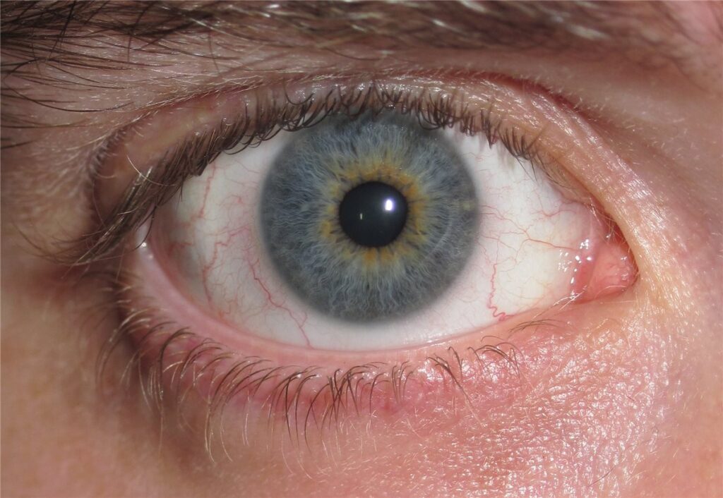
- Central heterochromia – a ring of different colour around the pupil within a single iris, often a normal variant
- Sectoral heterochromia – a wedge or patch of different colour within one iris, which can be a normal variant or, particularly when new, warrants closer evaluation
Congenital Causes
Simple Heterochromia
A benign, isolated finding present from birth, with no other ocular or systemic abnormality, likely reflecting normal variation in melanocyte distribution during iris development. This is by far the most common cause encountered in everyday practice and requires no investigation once other causes have been reasonably excluded by history and examination.
Horner Syndrome
Congenital Horner syndrome, whether from birth trauma or another perinatal cause, characteristically produces heterochromia with the affected iris remaining lighter in colour, since sympathetic innervation influences iris melanocyte development in early life. The affected eye also shows ptosis and miosis, and the combination of these three findings in an infant should prompt evaluation for an underlying cause, including imaging of the sympathetic chain in some cases.
Waardenburg Syndrome
A genetic disorder combining heterochromia (or strikingly pale, often blue, irides) with sensorineural hearing loss and characteristic white forelock and skin pigmentary changes, distinguished by its distinctive combination of features beyond the eye alone.
Congenital Horner-Associated Neuroblastoma
Congenital Horner syndrome with heterochromia occasionally signals an underlying neuroblastoma of the sympathetic chain, which is why any infant presenting with this combination warrants prompt paediatric evaluation and appropriate imaging rather than being assumed to be a benign isolated finding.
All-fit Slit-Lamp Adapter
Record and share exactly what you see at the slit lamp. One adapter fits any slit lamp or surgical microscope — and any smartphone.
From Choroida — the team behind this siteAcquired Causes
Chronic Anterior Uveitis
Longstanding inflammation, as seen in Fuchs heterochromic iridocyclitis, causes progressive iris stromal atrophy, typically leading to a lighter, mottled appearance of the affected eye, sometimes accompanied by fine, scattered keratic precipitates and, notably, without significant synechiae even after years of low-grade inflammation.
Iron Deposition (Siderosis)
A retained iron-containing intraocular foreign body can cause progressive iris discolouration, typically toward a rusty or heterochromic brown, along with other signs of siderosis bulbi affecting the lens and retina over time, making a careful trauma history relevant in any new, progressive heterochromia.
Iris or Ciliary Body Tumours
Melanoma, naevus, or other pigmented lesions of the iris or ciliary body can produce heterochromia, particularly when diffuse rather than localised, and this possibility is an important reason any newly acquired heterochromia deserves careful examination rather than reassurance by default.
Medication-Induced
Topical prostaglandin analogues, used in glaucoma treatment, cause progressive darkening of the iris in a substantial proportion of treated patients, typically most noticeable in eyes with mixed-colour irides, and this is a well-recognised, expected side effect rather than a sign of underlying pathology.
Evaluation
The single most important historical question is whether the colour difference has been present since infancy or early childhood, in which case a benign congenital cause is far more likely, or whether it has appeared or changed recently in an adult, which shifts concern meaningfully toward an acquired cause.
Slit-lamp examination should assess for keratic precipitates, anterior chamber cells, iris stromal atrophy or nodularity, and any discrete mass, and gonioscopy is useful when a tumour or foreign body is suspected.
A dilated fundus examination and, where a tumour is suspected, ultrasound biomicroscopy or anterior segment OCT help characterise a suspicious lesion further, and old photographs, including a driving licence or passport photo, can be genuinely useful for establishing how long a colour difference has actually been present when the history alone is unclear.
Differential Diagnosis Summary
- Simple congenital heterochromia – stable since birth, no other findings
- Horner syndrome – ptosis and miosis on the lighter side, congenital or acquired
- Fuchs heterochromic iridocyclitis – fine keratic precipitates, iris atrophy, absence of synechiae despite chronic inflammation
- Siderosis from a retained foreign body – relevant trauma history, progressive change
- Iris melanoma or naevus – a visible mass or diffuse pigment change, particularly if new or changing
- Prostaglandin analogue use – clear medication history, gradual darkening over months
Management
Isolated congenital heterochromia with no other findings requires no treatment or ongoing monitoring beyond routine eye care.
Congenital Horner syndrome warrants paediatric assessment to exclude an underlying neuroblastoma or other cause, coordinated with paediatrics and, where indicated, imaging of the neck and chest.
Acquired causes are managed according to the underlying diagnosis: uveitis treatment for Fuchs heterochromic iridocyclitis, removal of a retained foreign body where feasible in siderosis, and referral to ocular oncology for any suspicious pigmented lesion.
Prognosis
Congenital, isolated heterochromia carries an excellent prognosis and no visual consequence beyond its cosmetic appearance.
Outcome in acquired heterochromia depends entirely on the underlying cause, ranging from benign and stable in medication-induced darkening to potentially sight- or life-threatening in the case of an underlying tumour or unrecognised neuroblastoma.
The practical value of a systematic approach to heterochromia is that it reliably separates the large majority of patients who need only reassurance from the small minority who need urgent further investigation, without requiring extensive testing in every case.


Document what you see
Two smartphone imaging tools built for everyday clinic use — one for the slit lamp, one for the fundus.
From Choroida — the team behind this siteReferences
- Bansal S, Barathi VA, Iwata D, Agrawal R. Optical coherence tomography in the diagnosis and management of uveitis. International Ophthalmology. 2018.
- Weiss JS, Bilezikian JP. Fuchs heterochromic iridocyclitis. Survey of Ophthalmology. 1988.
- George ND, Gonzalez G, Hoyt CS. Does Horner’s syndrome in infancy require investigation? British Journal of Ophthalmology. 1998.
- Heterochromia Iridis. EyeWiki, American Academy of Ophthalmology.
- Heterochromia. StatPearls, NCBI Bookshelf.
Test yourself
A few questions straight from this article.
-
Which historical question matters most when assessing iris heterochromia?
Colour present since infancy points strongly to a benign congenital cause, while a difference that has appeared or changed in an adult shifts concern toward an acquired cause. -
What defines central heterochromia of the iris?
Central heterochromia is a ring of different colour around the pupil within a single iris and is often a normal variant. -
How does the iris appear in congenital Horner syndrome?
Sympathetic innervation influences iris melanocyte development early in life, so the affected iris stays lighter, alongside ptosis and miosis on the same side. -
Which condition must be excluded in an infant with congenital Horner syndrome and heterochromia?
This combination occasionally signals a neuroblastoma of the sympathetic chain, so it warrants prompt paediatric assessment and imaging rather than reassurance. -
Which features accompany heterochromia in Waardenburg syndrome?
Waardenburg syndrome combines heterochromia or strikingly pale irides with sensorineural hearing loss and pigmentary changes including a white forelock. -
Which finding characterises Fuchs heterochromic iridocyclitis?
Chronic low-grade inflammation produces progressive iris stromal atrophy with a lighter mottled iris and fine scattered keratic precipitates, notably without significant synechiae. -
Which history is relevant when iris heterochromia is progressive and rusty brown?
A retained iron-containing intraocular foreign body causes progressive rusty iris discolouration with other features of siderosis bulbi, making a trauma history essential. -
Which topical glaucoma medication causes progressive darkening of the iris?
Topical prostaglandin analogues darken the iris progressively in a substantial proportion of treated patients, most noticeably in mixed-colour irides, and this is an expected side effect. -
How should a newly acquired diffuse iris pigment change be managed?
Melanoma, naevus and other pigmented lesions can produce heterochromia, particularly when diffuse, so any suspicious pigmented lesion goes to ocular oncology. -
Which practical aid helps establish how long iris heterochromia has been present?
Old photographs, including a driving licence or passport photo, can settle how long a colour difference has actually been present when the history is unclear.

Writes and reviews the clinical articles on Educate. Every post is checked against peer-reviewed sources before it goes live.


