
Cavernous sinus thrombosis is thrombosis of the venous sinus that drains the orbit, sinuses, and much of the face, and it is one of the few ophthalmic emergencies where the eye findings are really a window onto a life-threatening intracranial process.
Before antibiotics, this condition was almost uniformly fatal. It remains dangerous today, and the eye findings, bilateral involvement in particular, are often what first raises the alarm.
Anatomy and Pathophysiology
The cavernous sinus receives venous drainage from the orbit via the superior and inferior ophthalmic veins, and from the face via the facial and angular veins, all of which are valveless.
This valveless drainage is the key vulnerability: infection from the face, sinuses, or orbit can spread directly backward into the cavernous sinus rather than being restricted to its site of origin, and infection reaching one cavernous sinus can spread to the other through connecting venous channels.
Once infected thrombus forms within the sinus, it obstructs venous outflow from the orbit and compresses the cranial nerves that pass through or alongside the sinus, specifically III, IV, V1, V2, and VI, producing the combination of orbital congestion and ophthalmoplegia that characterises the condition.
Aetiology
- Contiguous spread from sinusitis, particularly the ethmoid and sphenoid sinuses, given their close anatomical proximity to the cavernous sinus
- Facial infection, classically from the “danger triangle” of the face, where superficial facial veins communicate directly with the cavernous sinus via the ophthalmic veins
- Orbital cellulitis progressing posteriorly
- Odontogenic infection and mastoiditis, less common but recognised sources
- Staphylococcus aureus is the most frequently identified organism, though streptococcal species and, notably, invasive fungal infection with Mucor or Aspergillus species occur disproportionately in immunocompromised or diabetic patients and carry a particularly poor prognosis
All-fit Slit-Lamp Adapter
Record and share exactly what you see at the slit lamp. One adapter fits any slit lamp or surgical microscope — and any smartphone.
From Choroida — the team behind this siteClinical Presentation
The presentation typically begins unilaterally with headache, fever, and orbital or periorbital swelling, often following a preceding sinus, facial, or dental infection. Rapid progression to the fellow eye, over hours to a day or two, is the finding that should immediately raise concern for cavernous sinus thrombosis rather than simple orbital cellulitis.
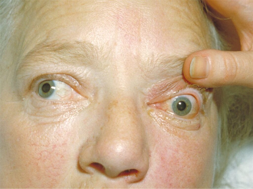
Proptosis, chemosis, and ophthalmoplegia are typically present, with the sixth nerve frequently affected first given its course directly through the sinus rather than along its wall. Ptosis and a dilated, poorly reactive pupil can occur from third nerve involvement, and periorbital and facial sensory loss can reflect involvement of the first and second trigeminal divisions.
Systemic signs of sepsis, including high fever and marked malaise, are usually present and reflect the severity of the underlying infection, and reduced consciousness or other neurological signs suggest intracranial extension.
Differential Diagnosis
- Orbital cellulitis without cavernous sinus involvement, which lacks bilateral progression and the specific pattern of multiple cranial nerve involvement
- Carotid-cavernous fistula, presenting with pulsatile proptosis and a bruit rather than the septic, rapidly progressive picture of thrombosis
- Idiopathic orbital inflammation, generally unilateral and without the systemic sepsis or bilateral spread seen in cavernous sinus thrombosis
- Thyroid eye disease, chronic and bilateral from the outset rather than acute and initially unilateral
Bilateral involvement developing rapidly after a unilateral start is the single most important clue distinguishing cavernous sinus thrombosis from orbital cellulitis, and it should shift management from routine orbital imaging to an urgent search for sinus venous thrombosis specifically.
Diagnostic Evaluation
MRI with MR venography is the investigation of choice, both to confirm sinus thrombosis directly and to characterise any associated orbital or intracranial abscess. CT venography is a reasonable alternative where MRI is not immediately available.
Blood cultures should be obtained before starting antibiotics wherever possible, since identifying the causative organism guides both antimicrobial therapy and the decision to pursue antifungal cover in higher-risk patients.
Lumbar puncture may be considered once raised intracranial pressure has been excluded on imaging, if meningitis is a concern, though it should not delay urgent antimicrobial treatment.
Management
Antimicrobial Therapy
High-dose intravenous broad-spectrum antibiotics covering Staphylococcus aureus, including MRSA where locally relevant, streptococcal species, and anaerobes should be started empirically without waiting for culture results, given the speed at which this condition can progress.
Antifungal cover should be added promptly in diabetic or immunocompromised patients, or where there is any clinical suspicion of invasive fungal sinusitis, since mucormycosis in particular can be rapidly fatal if the diagnosis is missed.
Anticoagulation
Anticoagulation remains debated but is used in many centres to limit thrombus propagation, balanced against the bleeding risk in a patient who may also need urgent sinus or orbital surgery.
Surgical Source Control
Drainage of the underlying sinus infection or any associated abscess is indicated when a surgically accessible source is identified, and is particularly urgent in suspected invasive fungal disease, where aggressive surgical debridement alongside antifungal therapy is central to survival.
Supportive Care
Management in a high-dependency or intensive care setting with close neurological and ophthalmic monitoring is appropriate given the risk of rapid deterioration, and close multidisciplinary involvement from neurosurgery, otolaryngology, infectious disease, and ophthalmology is standard.
Complications
- Permanent vision loss from optic nerve compression, central retinal artery or vein occlusion, or secondary glaucoma
- Meningitis, cerebral abscess, and other forms of intracranial extension
- Pituitary dysfunction from thrombosis extending to compromise pituitary blood supply
- Death, historically the expected outcome and still a real risk today, particularly with delayed diagnosis or invasive fungal disease
Prognosis
Mortality has fallen substantially with modern antimicrobial therapy but remains meaningfully elevated compared with most other orbital and periorbital infections, and is markedly worse in cases caused by invasive fungal infection.
Among survivors, cranial nerve palsies often improve over weeks to months as the underlying sinus thrombosis resolves, though visual outcome depends heavily on how much ischaemic damage occurred to the optic nerve or retina before treatment began.
Speed of diagnosis and treatment initiation is the dominant factor in outcome, which is precisely why bilateral progression of what initially looked like unilateral orbital cellulitis needs to be recognised and acted on immediately rather than managed as a routine, slowly escalating infection.


Document what you see
Two smartphone imaging tools built for everyday clinic use — one for the slit lamp, one for the fundus.
From Choroida — the team behind this siteReferences
- Southwick FS, Richardson EP, Swartz MN. Septic thrombosis of the dural venous sinuses. Medicine. 1986.
- Ebright JR, Pace MT, Niazi AF. Septic thrombosis of the cavernous sinuses. Archives of Internal Medicine. 2001.
- DiNubile MJ. Septic thrombosis of the cavernous sinuses. Archives of Neurology. 1988.
- Cavernous Sinus Thrombosis. EyeWiki, American Academy of Ophthalmology.
- Cavernous Sinus Thrombosis. StatPearls, NCBI Bookshelf.
Test yourself
A few questions straight from this article.
-
Which anatomical feature allows facial infection to spread directly into the cavernous sinus?
The ophthalmic, facial and angular veins draining into the cavernous sinus are valveless, so infection can travel backward into the sinus instead of remaining at its site of origin. -
In cavernous sinus thrombosis, which cranial nerve is frequently affected first?
The sixth nerve runs directly through the cavernous sinus rather than along its wall, which is why it is frequently the first to be involved. -
Which organism is most frequently identified in septic cavernous sinus thrombosis?
Staphylococcus aureus is the organism most often isolated, with streptococcal species and invasive fungal infection accounting for many of the remainder. -
Which feature most strongly suggests cavernous sinus thrombosis rather than simple orbital cellulitis?
Bilateral involvement developing rapidly after a unilateral onset is the single most important clue, and it should trigger an urgent search for sinus venous thrombosis. -
Which presentation points to carotid-cavernous fistula rather than cavernous sinus thrombosis?
A carotid-cavernous fistula produces pulsatile proptosis and a bruit, rather than the septic, rapidly progressive picture seen in cavernous sinus thrombosis. -
Which investigation is the study of choice to confirm cavernous sinus thrombosis?
MRI with MR venography confirms the sinus thrombosis directly and characterises any associated orbital or intracranial abscess; CT venography is a reasonable alternative. -
In cavernous sinus thrombosis, which patients should receive prompt antifungal cover?
Antifungal cover is added promptly in diabetic or immunocompromised patients because mucormycosis can be rapidly fatal if the diagnosis is missed. -
Why is anticoagulation used in many centres for cavernous sinus thrombosis?
Anticoagulation remains debated, but many centres use it to limit thrombus propagation, weighed against bleeding risk in a patient who may need urgent surgery. -
Which endocrine complication can follow cavernous sinus thrombosis?
Thrombosis extending to compromise the pituitary blood supply can leave lasting pituitary dysfunction. -
What is the typical course of cranial nerve palsies in survivors of cavernous sinus thrombosis?
Among survivors, cranial nerve palsies often improve over weeks to months as the underlying sinus thrombosis resolves.

Writes and reviews the clinical articles on Educate. Every post is checked against peer-reviewed sources before it goes live.


