
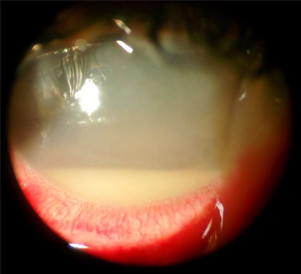
Acute postoperative endophthalmitis is intraocular infection presenting within roughly the first one to two weeks after intraocular surgery, most commonly cataract surgery given how frequently it is performed.
Its absolute incidence is low, but the consequences of a missed or delayed diagnosis are severe enough that any patient with unexpected pain, redness, or reduced vision in the days after surgery needs to be seen and assessed the same day, not the next available routine slot.
Pathophysiology
Bacteria are introduced into the eye at the time of surgery, most often from the patient’s own periocular and conjunctival flora, despite standard perioperative antisepsis with povidone-iodine.
Once inside the eye, organisms proliferate in the vitreous cavity, triggering an intense inflammatory response. This inflammatory response, as much as the infection itself, is what drives much of the tissue damage and visual loss seen in severe cases.
Staphylococcus epidermidis and other coagulase-negative staphylococci account for the majority of culture-positive cases, generally causing a less fulminant course. Staphylococcus aureus and streptococcal species are less common but tend to produce more rapidly destructive disease.
Risk Factors
- Posterior capsule rupture or vitreous loss during surgery, a well-established risk factor across multiple large surgical outcome studies
- Clear corneal incisions, particularly if not well-constructed or if wound integrity is compromised postoperatively
- Prolonged surgical time and the use of multiple instruments or repeated entries into the eye
- Diabetes and other conditions associated with impaired wound healing or immune response
- Use of silicone rather than acrylic intraocular lenses in some studies, though this association is less consistently reproduced than the surgical risk factors above
- Contamination of irrigation solutions or improperly sterilised instruments, an uncommon but recognised cause of cluster outbreaks
Fundus Explorer Pro
Photograph the retinal findings described here with the phone already in your pocket — 22 D optics and built-in illumination in one handheld unit.
From Choroida — the team behind this siteClinical Presentation
Pain, typically more severe than expected for routine postoperative recovery, is a common and important symptom, though a subset of patients, particularly with less virulent organisms, have relatively little pain.
Reduced vision, redness, and increasing lid swelling over the days following surgery should raise concern, especially when these findings represent a clear worsening rather than the expected gradual improvement of normal postoperative recovery.
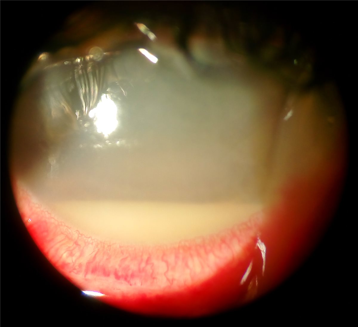
On examination, a hypopyon, layered white cells in the anterior chamber, is a hallmark finding, though its absence does not exclude the diagnosis, particularly early in the course. Marked anterior chamber reaction, corneal oedema, and vitreous inflammation obscuring the fundus view are typically present.
A relative afferent pupillary defect or absent red reflex indicates more severe involvement and a worse visual prognosis, and should be documented at initial assessment as a baseline for tracking response to treatment.
Differential Diagnosis
- Toxic anterior segment syndrome, a sterile inflammatory reaction presenting within the first 24 to 48 hours, typically without significant pain and often responding rapidly to topical steroids rather than progressing
- Retained lens fragments causing a sterile inflammatory reaction, distinguished by the surgical history and typically a less severe, more indolent course
- Uveitis-glaucoma-hyphema syndrome in pseudophakic eyes, presenting later and without the acute infective features
- Simple postoperative inflammation, which is expected to improve steadily rather than worsen, and lacks a hypopyon or severe pain
Distinguishing infectious endophthalmitis from toxic anterior segment syndrome is the most clinically important differential, since the two require essentially opposite initial management, and the timing of onset relative to surgery is one of the more useful discriminating clues.
Diagnostic Evaluation
Diagnosis is primarily clinical, and treatment should not be delayed while waiting for microbiology results in a patient with a convincing presentation.
Vitreous and aqueous samples are obtained at the time of intravitreal injection, both for culture and Gram stain, to guide any later adjustment of antibiotic therapy once results are available.
B-scan ultrasonography can help assess the extent of vitreous involvement when the view of the fundus is too poor for direct examination, and is useful for planning whether early vitrectomy may be needed.
Management
Immediate Treatment
Intravitreal antibiotics, typically a combination covering both Gram-positive and Gram-negative organisms, are administered emergently once the diagnosis is suspected, alongside vitreous and aqueous sampling for culture.
The Role of Vitrectomy
The Endophthalmitis Vitrectomy Study established that immediate pars plana vitrectomy improves outcomes specifically in eyes presenting with light perception vision only, while eyes with better presenting vision do just as well with intravitreal antibiotic injection alone as an initial approach, without needing immediate vitrectomy.
This distinction by presenting visual acuity remains the basis for the initial treatment decision in most cases, though vitrectomy is also considered if a patient fails to respond adequately to intravitreal antibiotics over the following days.
Adjunctive Therapy
Topical and sometimes systemic antibiotics are used as adjuncts, alongside topical corticosteroids once infection is controlled, to manage the accompanying inflammatory response. Topical cycloplegics provide symptomatic comfort and help prevent posterior synechiae.
Prognosis
Outcomes depend heavily on the infecting organism and how quickly treatment was started. Coagulase-negative staphylococcal infections generally carry the best visual prognosis, while more virulent organisms such as Staphylococcus aureus, streptococci, or Gram-negative organisms are associated with worse outcomes despite prompt treatment.
Presenting visual acuity is one of the strongest predictors of final outcome, which is exactly why it is used to guide the initial treatment decision between injection alone and immediate vitrectomy.
Delay between symptom onset and treatment initiation is consistently associated with worse visual outcomes across the literature, reinforcing why same-day assessment of any patient with concerning postoperative symptoms is the standard that protects vision in this condition.


Document what you see
Two smartphone imaging tools built for everyday clinic use — one for the slit lamp, one for the fundus.
From Choroida — the team behind this siteReferences
- Endophthalmitis Vitrectomy Study Group. Results of the Endophthalmitis Vitrectomy Study. Archives of Ophthalmology. 1995.
- Kessel L, Flesner P, Andresen J, et al. Antibiotic prevention of postcataract endophthalmitis: a systematic review and meta-analysis. Acta Ophthalmologica. 2015.
- Han DP, Wisniewski SR, Wilson LA, et al. Spectrum and susceptibilities of microbiologic isolates in the Endophthalmitis Vitrectomy Study. American Journal of Ophthalmology. 1996.
- Acute Postoperative Endophthalmitis. EyeWiki, American Academy of Ophthalmology.
- Endophthalmitis. StatPearls, NCBI Bookshelf.
Test yourself
A few questions straight from this article.
-
Acute postoperative endophthalmitis typically presents within what interval after intraocular surgery?
The acute form is defined by intraocular infection appearing in approximately the first one to two weeks after intraocular surgery, most commonly cataract surgery. -
In acute postoperative endophthalmitis, where do the causative bacteria most often originate?
Organisms are usually introduced at surgery from the patient's own periocular and conjunctival flora, despite standard antisepsis with povidone-iodine. -
Which organisms account for the majority of culture-positive acute postoperative endophthalmitis cases?
Staphylococcus epidermidis and other coagulase-negative staphylococci make up most culture-positive cases and generally follow a less fulminant course. -
Which intraoperative event is a well-established risk factor for acute postoperative endophthalmitis?
Posterior capsule rupture or vitreous loss during surgery is a risk factor established across multiple large surgical outcome studies. -
Which anterior segment finding is the hallmark sign of acute postoperative endophthalmitis?
Layered white cells in the anterior chamber form the hallmark hypopyon, although its absence does not exclude the diagnosis, particularly early on. -
Which feature best distinguishes toxic anterior segment syndrome from acute postoperative endophthalmitis?
Toxic anterior segment syndrome is a sterile reaction appearing within 24 to 48 hours, typically painless, and it responds rapidly to topical steroids rather than progressing. -
In the Endophthalmitis Vitrectomy Study, which eyes benefited from immediate pars plana vitrectomy?
Immediate vitrectomy improved outcomes specifically in eyes presenting with light perception only; eyes with better presenting vision did equally well with intravitreal antibiotics alone. -
When the fundus view is obscured in suspected endophthalmitis, which test assesses vitreous involvement?
B-scan ultrasonography shows the extent of vitreous involvement when the media are too hazy for direct examination, and helps decide whether early vitrectomy is needed. -
Which presenting finding in acute postoperative endophthalmitis signals severe involvement and a worse visual prognosis?
A relative afferent pupillary defect or an absent red reflex marks more severe involvement and a worse prognosis, and should be documented as a treatment-response baseline. -
Which factor is consistently associated with worse visual outcomes in acute postoperative endophthalmitis?
Delay from symptom onset to treatment is consistently linked with worse vision across the literature, which is why any concerning postoperative symptom warrants same-day assessment.

Writes and reviews the clinical articles on Educate. Every post is checked against peer-reviewed sources before it goes live.


