
Strabismus surgery adjusts the position of one or more extraocular muscles to correct ocular misalignment, most commonly by weakening an overacting muscle through recession or strengthening an underacting one through resection.
The underlying mechanics are simple to describe and considerably harder to execute precisely, since the amount of correction achieved depends on measurements, surgical technique, and a certain amount of biological variability that no formula fully accounts for.
Surgical Principles
Recession weakens a muscle by detaching it from its original insertion and reattaching it further back on the globe, reducing its mechanical pull. Resection strengthens a muscle by removing a segment of it and reattaching the shortened muscle at its original insertion, increasing its effective pull.
The amount of recession or resection is calculated from the preoperative angle of deviation, measured in prism dioptres, using standard surgical dosage tables developed from decades of outcome data. These tables provide a starting point, but experienced surgeons adjust based on the specific pattern of strabismus and any prior surgery.
Combining recession of one muscle with resection of its antagonist, most classically medial rectus recession paired with lateral rectus resection for exotropia, or the reverse for esotropia, is a common approach for correcting horizontal strabismus in a single eye.
Indications
- Constant or significant intermittent strabismus not adequately controlled by non-surgical measures such as glasses or prisms
- Strabismus causing diplopia, confirmed to be relievable by the planned surgical correction
- Significant strabismus affecting binocular function or causing psychosocial impact from the visible misalignment
- Restrictive or paretic strabismus following trauma, thyroid eye disease, or cranial nerve palsy, once the deviation has stabilised
- As an adjunct in conditions such as Duane syndrome, where surgery improves head posture and alignment even though it cannot restore normal muscle function
All-fit Slit-Lamp Adapter
Record and share exactly what you see at the slit lamp. One adapter fits any slit lamp or surgical microscope — and any smartphone.
From Choroida — the team behind this sitePreoperative Assessment
Accurate measurement of the deviation at distance and near, in different gaze positions, is the foundation of surgical planning, since the pattern of the strabismus, not just its overall magnitude, determines which muscles are operated on.
Assessment for any restrictive component with forced duction testing distinguishes a purely paretic deviation from one with a mechanical restriction, which is managed differently and can change the surgical plan substantially.
Sensory status, including presence and depth of binocular fusion and any suppression, informs both the likelihood of achieving binocular single vision after surgery and the discussion of realistic expectations with the patient or family.
Surgical Technique
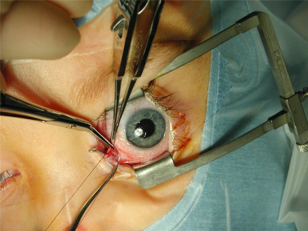
The conjunctiva and Tenon capsule are opened to expose the target muscle, which is isolated on a muscle hook and secured with a suture before being disinserted from the sclera.
For recession, the muscle is reattached posterior to its original insertion at a measured distance; for resection, a measured segment is excised before reattachment at the original insertion site.
Adjustable suture techniques, where the muscle is secured with a slipknot that can be modified in the early postoperative period based on measured alignment, are used in selected adult patients to fine-tune the result before it is finally fixed.
Complications
- Overcorrection or undercorrection, the most common reason for a second surgery, reflecting the inherent variability in how individual eyes respond to a given amount of muscle adjustment
- Diplopia, usually transient as the visual system adapts to the new alignment, but occasionally persistent, particularly in adults with long-standing suppression before surgery
- Scleral perforation during suture placement, rare with careful technique but a recognised risk given the thinness of the sclera at typical muscle insertion sites
- Anterior segment ischaemia, a rare but serious complication from disrupting the anterior ciliary vessels, more of a concern when multiple muscles are operated on in the same eye at once
- Infection or conjunctival scarring, uncommon with modern technique and perioperative care
The risk of anterior segment ischaemia is specifically why surgeons are cautious about operating on more than two rectus muscles of the same eye in a single procedure, staging further surgery when more extensive correction is needed.
Postoperative Course
Mild discomfort, redness, and swelling are expected in the days following surgery and generally settle within one to two weeks. Diplopia in the early postoperative period is common as the brain adjusts to the new alignment and usually resolves on its own.
Final alignment is typically assessed at follow-up visits over the following weeks to months, since some drift in position, in either direction, can occur during healing before the result stabilises.
Prognosis
Most patients achieve good functional and cosmetic alignment after strabismus surgery, though a meaningful proportion require a second procedure at some point, either for undercorrection, overcorrection, or drift over subsequent years, particularly in complex or paralytic strabismus.
Restoration of binocular fusion depends heavily on preoperative sensory status. Patients with good preoperative fusion potential have the best chance of regaining stereopsis after alignment is corrected, while those with long-standing suppression or amblyopia may achieve cosmetic realignment without full binocular function.
Surgery for restrictive or paretic strabismus, including in conditions like Duane syndrome or after cranial nerve palsy, improves alignment and head posture but does not restore normal muscle function where the underlying nerve or mechanical problem persists.


Document what you see
Two smartphone imaging tools built for everyday clinic use — one for the slit lamp, one for the fundus.
From Choroida — the team behind this siteReferences
- Wright KW, Strube YNJ. Color Atlas of Strabismus Surgery. Springer.
- Helveston EM. Complications of strabismus surgery. Current Opinion in Ophthalmology. 2009.
- American Academy of Ophthalmology. Pediatric Eye Evaluations and Esotropia and Exotropia Preferred Practice Pattern.
- Strabismus Surgery. EyeWiki, American Academy of Ophthalmology.
- Strabismus Surgery Techniques. StatPearls, NCBI Bookshelf.
Test yourself
A few questions straight from this article.
-
How does recession weaken an extraocular muscle in strabismus surgery?
The muscle is detached from its original insertion and moved posteriorly, which reduces its mechanical pull on the eye. -
How does resection strengthen an extraocular muscle?
Shortening the muscle increases its effective pull while the insertion site stays where it was. Recession does the opposite by moving the insertion back. -
What is the amount of recession or resection calculated from?
Standard surgical dosage tables built from decades of outcome data convert the measured angle into millimetres of surgery, then the surgeon adjusts for the specific pattern and any prior operations. -
When is surgery undertaken for paretic strabismus following a cranial nerve palsy?
The same applies to restrictive strabismus after trauma or thyroid eye disease. Operating on a still-changing deviation invites an inaccurate result. -
What does forced duction testing add to preoperative strabismus assessment?
A mechanical restriction is managed differently from a purely paretic deviation, and finding one can change the surgical plan substantially. -
What is the purpose of an adjustable suture in strabismus surgery?
The muscle is secured with a slipknot that can be modified once alignment has been measured postoperatively. The technique is used in selected adult patients. -
What is the most common reason for a second strabismus operation?
Individual eyes respond variably to a given amount of muscle adjustment, and no dosage formula fully accounts for that biological variation. -
Why do surgeons avoid operating on more than two rectus muscles of one eye at once?
Disrupting multiple anterior ciliary vessels in one eye is the mechanism. More extensive correction is staged across separate procedures instead. -
How should diplopia in the first days after strabismus surgery be regarded?
It can occasionally persist, particularly in adults with long-standing suppression before surgery. Alignment itself is assessed over weeks to months as healing settles. -
What most determines whether binocular fusion is regained after strabismus surgery?
Good preoperative fusion potential gives the best chance of stereopsis after alignment. Long-standing suppression or amblyopia may leave cosmetic realignment without binocular function.

Writes and reviews the clinical articles on Educate. Every post is checked against peer-reviewed sources before it goes live.


