
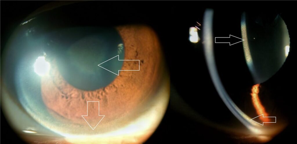
A Kayser-Fleischer ring is a golden-brown to greenish deposit of copper at the periphery of Descemet membrane, and it is one of the few ocular signs that can point directly to a specific systemic diagnosis: Wilson disease.
Its presence in a young patient with unexplained liver disease or new neuropsychiatric symptoms is a genuinely useful bedside finding, and one that can be picked up at the slit lamp before serum copper studies come back.
What Causes the Ring?
Wilson disease is an autosomal recessive disorder of copper metabolism caused by mutations in ATP7B, a gene encoding a copper-transporting ATPase in the liver.
Loss of this transporter impairs biliary copper excretion, causing copper to accumulate first in the liver and, once hepatic storage capacity is exceeded, in other tissues including the brain, kidneys, and eye.
In the eye, copper deposits in the peripheral corneal stroma, specifically at the level of Descemet membrane, forming a ring that is usually most visible at the superior and inferior limbus before it extends circumferentially as the disease progresses.
Clinical Significance
A Kayser-Fleischer ring is present in the large majority of patients with neurological Wilson disease, but in a smaller proportion of those with liver disease alone and no neurological involvement.
Its absence, particularly in a patient with predominantly hepatic disease, does not exclude Wilson disease. Its presence, conversely, is a strong pointer toward the diagnosis in the right clinical context, though it is not entirely specific to Wilson disease on its own.
All-fit Slit-Lamp Adapter
Record and share exactly what you see at the slit lamp. One adapter fits any slit lamp or surgical microscope — and any smartphone.
From Choroida — the team behind this siteExamination Findings
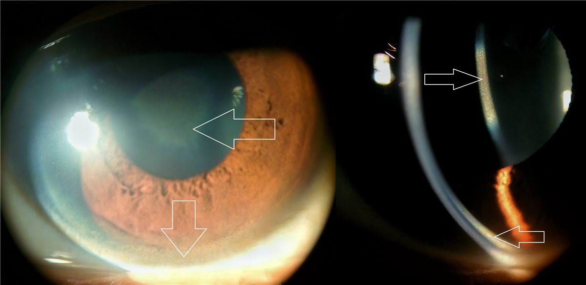
Early or subtle rings can be missed on routine slit-lamp examination with white light, particularly in patients with dark irides, and are better seen with the beam directed obliquely at the limbus.
Gonioscopy or slit-lamp examination by an experienced examiner detects rings that are easily missed on cursory inspection, and formal ophthalmic assessment is recommended whenever Wilson disease is being actively considered, rather than relying on a brief penlight look.
The ring itself does not affect vision and causes no ocular symptoms; it is purely a diagnostic marker rather than a source of visual complaint.
Other Ocular Findings in Wilson Disease
- Sunflower cataract – a distinctive central, petal-like copper deposit in the anterior and posterior lens capsule, seen in a smaller subset of patients, also visually asymptomatic in most cases
- Rarely, copper deposition has been described affecting other ocular structures, though the corneal ring and sunflower cataract are by far the most clinically recognised findings
Differential Diagnosis
- Chalcosis, copper deposition from a retained intraocular copper-containing foreign body, distinguished by a clear trauma history and typically a more central rather than peripheral pattern
- Corneal arcus, a lipid deposit that can superficially resemble a limbal ring but is whitish-grey rather than golden-brown and unrelated to copper metabolism
- Other causes of chronic cholestatic liver disease with copper retention, such as primary biliary cholangitis, which can very rarely produce a similar corneal ring through a different mechanism of copper accumulation
Diagnostic Evaluation of Wilson Disease
Serum ceruloplasmin is typically low, and 24-hour urinary copper excretion is typically elevated, though neither test alone is fully sensitive or specific, and results can be equivocal, particularly in early or hepatic-predominant disease.
Liver biopsy with quantitative hepatic copper measurement remains the most definitive test when the diagnosis is uncertain from serum and urine studies alone. Genetic testing for ATP7B mutations is increasingly used, particularly in families where a mutation has already been identified in an affected relative.
The combination of a Kayser-Fleischer ring with low ceruloplasmin and elevated urinary copper is highly suggestive of Wilson disease and is often sufficient to proceed with treatment without further invasive testing.
Management
Treatment of Wilson disease itself is medical, using copper chelation with agents such as penicillamine or trientine, or zinc salts to reduce intestinal copper absorption, managed by hepatology or a Wilson disease specialist.
The Kayser-Fleischer ring requires no separate ocular treatment. It typically fades or resolves gradually with effective systemic chelation therapy, and its regression over time is itself used as a useful, if imperfect, marker of treatment response.
Ongoing ophthalmic examination is reasonable during treatment, both to confirm ring resolution as a supportive sign of adequate copper control and to monitor for any changes in the sunflower cataract if present.
Prognosis
The ring itself carries no independent visual prognosis and does not threaten sight. The prognosis that matters is that of the underlying Wilson disease, which is generally good with early diagnosis and consistent treatment, but can progress to liver failure or severe neurological disability if missed or inadequately treated.
Regression of the Kayser-Fleischer ring with treatment is a reassuring sign of adequate systemic copper control, while a ring that fails to regress, or reappears after having resolved, should prompt reassessment of treatment adherence and efficacy.


Document what you see
Two smartphone imaging tools built for everyday clinic use — one for the slit lamp, one for the fundus.
From Choroida — the team behind this siteReferences
- Roberts EA, Schilsky ML. Diagnosis and treatment of Wilson disease: an update. Hepatology. 2008.
- Suvarna JC. Kayser-Fleischer ring. Journal of Postgraduate Medicine. 2008.
- European Association for the Study of the Liver. EASL Clinical Practice Guidelines: Wilson’s disease. Journal of Hepatology. 2012.
- Kayser-Fleischer Ring. EyeWiki, American Academy of Ophthalmology.
- Wilson Disease. StatPearls, NCBI Bookshelf.
Test yourself
A few questions straight from this article.
-
Where is the copper deposited in a Kayser-Fleischer ring?
The ring is a golden-brown to greenish copper deposit at the corneal periphery, at the level of Descemet membrane. It is one of the few ocular signs pointing straight to a systemic diagnosis. -
Which defect underlies copper accumulation in Wilson disease?
ATP7B encodes a copper-transporting ATPase in the liver. Losing it means copper builds up hepatically first, then spills into the brain, kidneys and eye once storage capacity is exceeded. -
What does a Kayser-Fleischer ring mean in a patient being assessed for Wilson disease?
The ring is present in the large majority of neurological Wilson disease but a smaller proportion of purely hepatic cases. A negative slit-lamp finding must not close the question. -
Where is a Kayser-Fleischer ring usually first visible at the slit lamp?
The ring starts superiorly and inferiorly, then extends circumferentially as disease progresses. Early rings are easily missed in dark irides unless the beam is angled obliquely at the limbus. -
How does a Kayser-Fleischer ring affect the patient's vision?
The ring is purely a diagnostic marker, never a source of visual complaint. Its whole value lies in what it signals about the liver and brain. -
Which other ocular finding is recognised in Wilson disease?
This is a central, petal-like copper deposit in the anterior and posterior lens capsule, seen in a smaller subset of patients and usually visually asymptomatic. -
How does corneal arcus differ from a Kayser-Fleischer ring at the slit lamp?
Arcus is a lipid deposit with no relation to copper metabolism, despite sitting in a similar limbal position. Colour is the discriminator at the slit lamp. -
Which test is most definitive when serum and urine studies leave Wilson disease uncertain?
Low ceruloplasmin and raised 24-hour urinary copper are neither fully sensitive nor specific and can be equivocal early on. Biopsy settles genuinely uncertain cases. -
How is Wilson disease itself treated once the diagnosis is made?
Penicillamine or trientine chelate copper, while zinc reduces intestinal absorption, under hepatology care. The ring itself needs no separate ocular treatment and fades with effective therapy. -
What does a Kayser-Fleischer ring that reappears after resolving suggest?
Ring regression is a useful, if imperfect, marker of adequate systemic copper control. A ring that fails to regress or returns points back at the treatment, not the eye.

Writes and reviews the clinical articles on Educate. Every post is checked against peer-reviewed sources before it goes live.


