
Duane retraction syndrome is a congenital eye movement disorder caused by absence or maldevelopment of the abducens nerve nucleus, with the lateral rectus instead receiving aberrant innervation from a branch of the oculomotor nerve.
It belongs to a group of conditions called congenital cranial dysinnervation disorders, where the problem lies in the wiring of the nerve, not in the muscle itself.
The eye movement pattern it produces is distinctive enough that most cases can be recognised on examination alone, without imaging.
Pathophysiology
In normal development, the abducens nucleus sends axons to the lateral rectus muscle, allowing abduction. In Duane syndrome, this nucleus is absent or hypoplastic.
The lateral rectus instead becomes innervated by an aberrant branch of the oculomotor nerve, which normally supplies the medial rectus.
Because both muscles now receive innervation from the same nerve, they co-contract on attempted adduction: the medial rectus pulls the eye inward as intended, but the lateral rectus contracts simultaneously instead of relaxing.
This co-contraction restricts adduction and causes the globe to retract into the orbit, narrowing the palpebral fissure. The mechanism explains every clinical feature of the syndrome.
Classification
Duane syndrome is divided into three types based on the pattern of horizontal movement restriction.
- Type I – limited abduction with normal or only mildly limited adduction; the most common form
- Type II – limited adduction with normal or only mildly limited abduction
- Type III – limited abduction and adduction, reflecting co-contraction affecting both directions
All three types share the same underlying mechanism of aberrant innervation; the difference lies in which muscle group is affected by the abnormal co-contraction, and to what degree.
All-fit Slit-Lamp Adapter
Record and share exactly what you see at the slit lamp. One adapter fits any slit lamp or surgical microscope — and any smartphone.
From Choroida — the team behind this siteClinical Presentation
Most children are brought in because of a noticeable turn of the head, which they adopt to maintain binocular single vision and avoid the restricted gaze position.
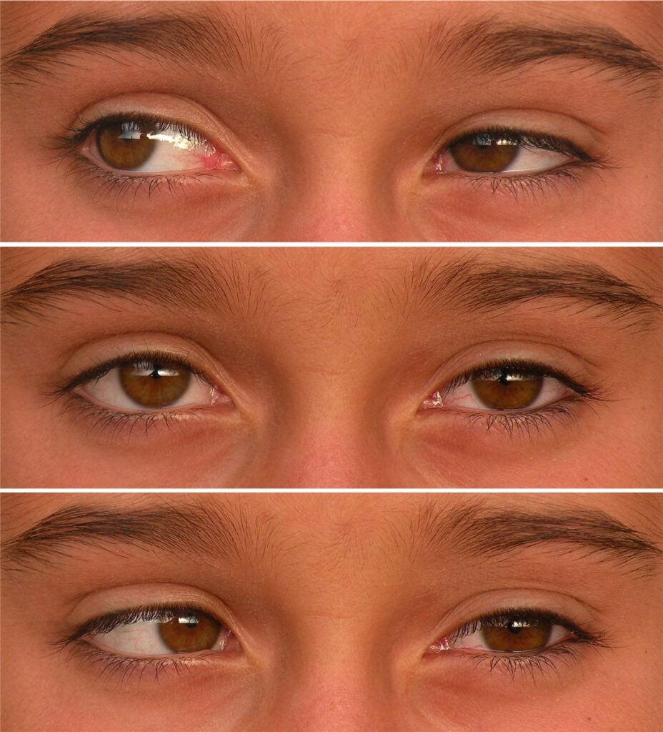
On examination, the hallmark sign is narrowing of the palpebral fissure and globe retraction on attempted adduction, with widening of the fissure on abduction. This retraction is what gives the syndrome its name and is usually obvious once looked for.
An upshoot or downshoot of the eye can occur on adduction in some patients, related to the abnormal path the co-contracting muscles take as they pull against each other. Strabismus, usually esotropia in type I, may be present in primary gaze, though many patients are only mildly misaligned or orthophoric when looking straight ahead.
Associated Findings
Duane syndrome occurs in isolation in most cases, but it is associated with other conditions often enough to warrant a broader look at the patient.
- Skeletal anomalies, including vertebral and limb defects, as seen in Okihiro syndrome
- Hearing loss, seen in Duane-radial ray syndrome and related conditions
- Other ocular anomalies, including microphthalmia and heterochromia in some cases
- Goldenhar syndrome, in which Duane syndrome can be one component of a broader craniofacial spectrum
A child with newly diagnosed Duane syndrome and any additional dysmorphic features or hearing concerns should be assessed more broadly rather than treated as an isolated ophthalmic finding.
Differential Diagnosis
- Sixth nerve palsy, which lacks the globe retraction and fissure narrowing characteristic of Duane syndrome
- Möbius syndrome, with bilateral facial and abducens palsy and a different pattern of restriction
- Congenital esotropia, distinguished by the absence of retraction and fissure narrowing on adduction
- Thyroid eye disease with restrictive myopathy, which presents later in life and lacks the congenital onset and retraction pattern
Globe retraction on adduction is the single most useful clinical sign separating Duane syndrome from a simple sixth nerve palsy, and it should be checked for specifically whenever abduction deficit is found in a child.
Evaluation
Diagnosis is clinical, based on the characteristic pattern of restriction and retraction described above. Forced duction testing, when performed, shows restriction consistent with co-contraction rather than a simple paretic pattern.
Neuroimaging is not routinely required for typical, isolated Duane syndrome, though it may be considered when the presentation is atypical or when other neurological findings raise concern.
Assessment for associated systemic anomalies is worthwhile in every newly diagnosed case, given the range of syndromes Duane syndrome can accompany. A basic hearing screen and general paediatric assessment are reasonable in a child with additional findings.
Management
Many children with Duane syndrome do not need surgery. Head turn alone, in the absence of significant strabismus in primary gaze or amblyopia, is often better tolerated than the risks of surgery would justify.
Surgery is considered for significant strabismus in primary position, a marked head turn causing functional or cosmetic concern, or significant upshoot or downshoot on adduction. Recession of the co-contracting muscles, most commonly the medial rectus, is used to reduce the abnormal pull rather than to restore normal abduction, which surgery cannot achieve since the underlying nerve supply cannot be corrected.
Amblyopia should be screened for and treated with standard occlusion therapy when present, independent of any decision about strabismus surgery.
Prognosis
Visual acuity is typically normal in Duane syndrome unless amblyopia develops from an untreated strabismus or significant anisometropia. With appropriate monitoring and amblyopia treatment when needed, visual outcomes are generally good.
Surgery, when performed, improves head posture and alignment in primary gaze but does not restore full range of eye movement, since the aberrant innervation itself is not correctable. Patients and families should be counselled about this limitation before surgery is considered.
Most patients with isolated Duane syndrome lead a normal life with normal binocular function in primary gaze, adapting well to whatever residual restriction remains after treatment.


Document what you see
Two smartphone imaging tools built for everyday clinic use — one for the slit lamp, one for the fundus.
From Choroida — the team behind this siteReferences
- Kekunnaya R, Negalur M. Duane retraction syndrome: causes, effects and management strategies. Clinical Ophthalmology. 2017.
- Yuksel D, Orban de Xivry JJ, Lefevre P. Review of the major findings about Duane retraction syndrome (DRS) leading to an updated form of classification. Vision Research. 2010.
- Engle EC. Genetic basis of congenital strabismus. Archives of Ophthalmology. 2007.
- Duane Retraction Syndrome. EyeWiki, American Academy of Ophthalmology.
- Duane Syndrome. StatPearls, NCBI Bookshelf.
Test yourself
A few questions straight from this article.
-
What is the underlying abnormality in Duane retraction syndrome?
The abducens nucleus is absent or hypoplastic, so the lateral rectus receives an aberrant branch of the oculomotor nerve instead. The muscle itself is normal; the wiring is not. -
Duane retraction syndrome belongs to which group of disorders?
These are conditions in which the fault lies in the development of the cranial nerve supply rather than in the muscle being innervated. -
Why does the globe retract on attempted adduction in Duane retraction syndrome?
Both muscles are driven by the same nerve, so the lateral rectus contracts instead of relaxing during adduction. The opposing pull drags the globe backwards and narrows the palpebral fissure. -
Which movement pattern characterises the most common type of Duane retraction syndrome?
This is type I, the commonest of the three forms. Types II and III show limited adduction, or restriction in both directions. -
What most often brings a child with Duane retraction syndrome to medical attention?
Children adopt the head turn to keep binocular single vision and avoid the restricted gaze position, and it is usually the parents' presenting complaint. -
Which sign separates Duane retraction syndrome from an isolated sixth nerve palsy?
A sixth nerve palsy limits abduction but produces no retraction. Retraction on adduction should be looked for specifically in any child with an abduction deficit. -
How is typical, isolated Duane retraction syndrome diagnosed?
The pattern of restriction and retraction is distinctive enough to make the diagnosis at the slit lamp or in the clinic. Neuroimaging is reserved for atypical cases or added neurological findings. -
Which associated finding warrants broader assessment in newly diagnosed Duane retraction syndrome?
Duane syndrome can accompany skeletal and hearing abnormalities, as in Duane-radial ray and Okihiro syndromes. A child with extra dysmorphic or hearing concerns needs assessment beyond the eye. -
When is surgery considered in a child with Duane retraction syndrome?
Significant upshoot or downshoot on adduction is a third indication. Many children do better with observation, since a tolerable head turn does not justify surgical risk. -
What can strabismus surgery realistically achieve in Duane retraction syndrome?
Recession of the co-contracting muscles reduces the abnormal pull, but the aberrant innervation cannot be corrected. Families should be counselled on this limit before surgery.

Writes and reviews the clinical articles on Educate. Every post is checked against peer-reviewed sources before it goes live.


