
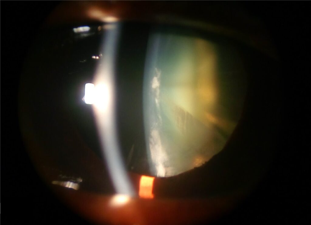
A cortical cataract is opacification of the peripheral lens cortex, classically producing wedge-shaped or spoke-like opacities that radiate from the lens equator toward the centre.
It is one of the three principal morphological patterns of age-related cataract, alongside nuclear sclerotic and posterior subcapsular cataract, and recognising the pattern on slit-lamp examination is useful well beyond simply describing what is seen, since it points toward a different symptom profile and a different rate of progression than the other two types.
Pathophysiology
The lens cortex is made up of lens fibres arranged in a highly organised radial pattern, and cortical cataract results from disruption of that architecture, most often attributed to osmotic stress within the lens fibres.
Water accumulates between disrupted fibre cells, forming clefts and vacuoles that scatter light, and this fluid accumulation is what gives cortical opacities their characteristic radial, spoke-like configuration rather than the diffuse, homogeneous change seen in nuclear sclerosis.
Oxidative damage to lens fibre membranes and proteins is thought to be a central mechanism, and this is consistent with the epidemiological association between cortical cataract and conditions or exposures that increase oxidative stress on the lens, including diabetes, prolonged ultraviolet exposure, and smoking.
Unlike nuclear sclerosis, which progresses in a fairly predictable, linear fashion with age, cortical cataract can progress unevenly, and individual spokes can enlarge, coalesce, or occasionally show partial resolution over a period of months, which makes serial documentation genuinely useful for tracking an individual patient’s disease course rather than assuming a uniform trajectory.
Risk Factors
- Advancing age, the dominant risk factor, though cortical cataract can develop at a younger age than nuclear sclerosis in some patients
- Diabetes mellitus, with cortical cataract specifically, rather than nuclear sclerosis, being the pattern most strongly associated with hyperglycaemia and osmotic lens changes
- Cumulative ultraviolet light exposure, which correlates with cortical cataract more strongly than with the other morphological types
- Smoking, associated with both nuclear and cortical cataract through separate oxidative mechanisms
- Prior ocular trauma or intraocular surgery, which can accelerate cortical change in the affected eye specifically
All-fit Slit-Lamp Adapter
Record and share exactly what you see at the slit lamp. One adapter fits any slit lamp or surgical microscope — and any smartphone.
From Choroida — the team behind this siteClinical Presentation
Symptoms depend heavily on whether the opacities involve the visual axis, and this is the key difference from nuclear sclerosis, where central involvement is essentially guaranteed by the nature of the disease.
Peripheral cortical spokes, which are common and often asymptomatic for a long period, may cause little more than mild glare or subtle contrast sensitivity loss, particularly noticeable in bright light or when driving at night against oncoming headlights.
Once spokes extend into the visual axis, patients typically report glare, especially with a bright point source, along with reduced contrast sensitivity, and monocular diplopia is occasionally described from light scattering unevenly through the irregular cortical opacity.
Because progression can be patchy and asymmetric between spokes, some patients report fluctuating vision, genuinely varying from day to day, which is different from the slow, steady decline more typical of nuclear sclerosis.
Examination Findings
On slit-lamp examination with retroillumination, cortical opacities appear as wedge-shaped or spoke-like white opacities radiating from the equator toward the centre of the lens, with clear lens material typically visible between individual spokes, at least in early to moderate disease.
As disease progresses, spokes can enlarge and coalesce, eventually obscuring the visual axis, and in advanced cases the entire cortex can become diffusely opacified.
Retinoscopy through a cortical cataract often shows an irregular, scissoring reflex, corresponding to the uneven distribution of the opacities across the pupil rather than the more uniform dulling seen with nuclear sclerosis.
A dilated examination is important specifically because peripheral spokes not visible through an undilated pupil can still be contributing meaningfully to glare symptoms that seem disproportionate to what is visible on undilated exam.
Differential Diagnosis
- Nuclear sclerotic cataract, with diffuse, central yellowing or browning of the lens nucleus rather than discrete peripheral spokes, and a more gradual, predictable myopic shift in refraction as it progresses
- Posterior subcapsular cataract, located just anterior to the posterior capsule, causing disproportionate glare and near-vision difficulty relative to its size, and classically associated with corticosteroid use or younger patient age compared with the other two types
- Traumatic cataract, which can produce a rosette-shaped opacity that superficially resembles cortical spokes but is distinguished by a clear history of trauma and typically a different, more stellate configuration
- Diabetic “snowflake” cataract, seen in poorly controlled diabetes, particularly in younger patients, and often more rapidly progressive than typical age-related cortical change
Management
No medical therapy has been shown to reverse or reliably slow cortical cataract progression, and management is therefore observational until visual symptoms become significant enough to warrant surgery.
Regular monitoring of visual acuity and, importantly, functional symptoms such as glare and contrast sensitivity is appropriate, since visual acuity on a standard chart, particularly in good lighting, can understate how much a patient is actually struggling in real-world conditions like night driving.
Cataract surgery, typically phacoemulsification with intraocular lens implantation, is indicated once visual symptoms meaningfully affect a patient’s daily function, and the threshold for proceeding should be guided by the patient’s own reported impact rather than a fixed visual acuity cutoff.
A relatively mild reduction in acuity accompanied by significant glare can be more functionally limiting than a lower acuity without glare. Patients with predominantly cortical cataract and disproportionate glare symptoms relative to their measured acuity are worth specifically counselling about this discrepancy, since it explains why their symptoms may feel more severe than a visual acuity number alone would suggest.
Prognosis
Cataract surgery for cortical cataract generally achieves excellent visual outcomes, similar to surgery for other cataract morphologies, since the underlying issue is a diseased crystalline lens rather than damage to the retina or optic nerve.
Progression rate before surgery is genuinely variable between patients and, at times, between eyes in the same patient, which makes fixed follow-up intervals somewhat less predictable to set than they are for nuclear sclerosis, and follow-up timing is better guided by the trajectory of an individual patient’s symptoms and findings than by a standard interval applied uniformly.
Once surgery is performed, outcomes are not meaningfully different from cataract surgery performed for any other morphological pattern, and the specific type of cataract removed has no bearing on long-term visual prognosis after a successful procedure.
Related Reading


Document what you see
Two smartphone imaging tools built for everyday clinic use — one for the slit lamp, one for the fundus.
From Choroida — the team behind this siteReferences
- Age-Related Eye Disease Study Research Group. Risk factors associated with age-related nuclear and cortical cataract. Ophthalmology. 2001.
- West S, Duncan DD, Munoz B, et al. Sunlight exposure and risk of lens opacities in a population-based study: the Salisbury Eye Evaluation project. JAMA. 1998.
- Michael R, Bron AJ. The ageing lens and cataract: a model of normal and pathological ageing. Philosophical Transactions of the Royal Society B. 2011.
- Cortical Cataract. EyeWiki, American Academy of Ophthalmology.
- Cataract. StatPearls, NCBI Bookshelf.
Test yourself
A few questions straight from this article.
-
Which lens appearance defines a cortical cataract?
Cortical cataract is opacification of the peripheral lens cortex, forming wedge-shaped or spoke-like opacities that radiate from the equator toward the centre. It is one of the three principal morphological patterns of age-related cataract. -
What process is held responsible for the radial spoke pattern in cortical cataract?
Osmotic stress disrupts the radial architecture of the lens fibres, and water collecting between them forms clefts and vacuoles that scatter light. This fluid accumulation gives the opacities their spoke-like configuration. -
Which systemic condition is most strongly associated with the cortical pattern of cataract specifically?
Cortical cataract, rather than nuclear sclerosis, is the pattern most strongly linked to hyperglycaemia and the resulting osmotic lens changes. Cumulative ultraviolet exposure and smoking are the other notable oxidative risk factors. -
What chiefly determines whether a cortical cataract is symptomatic?
Peripheral spokes are common and often asymptomatic for a long period. This is the key difference from nuclear sclerosis, where central involvement is guaranteed by the nature of the disease. -
Why do some patients with cortical cataract report vision that fluctuates from day to day?
Individual spokes can enlarge or coalesce unevenly, so vision genuinely varies between days. This differs from the slow, steady decline more typical of nuclear sclerosis. -
What does retinoscopy through a cortical cataract characteristically show?
The scissoring reflex reflects the uneven distribution of opacities across the pupil. Nuclear sclerosis instead produces a more uniform dulling of the reflex. -
Why is dilated examination important in a patient with suspected cortical cataract?
Spokes hidden behind an undilated pupil can contribute meaningfully to glare. This explains symptoms that otherwise seem disproportionate to what the undilated examination shows. -
Which features identify nuclear sclerotic rather than cortical cataract?
Nuclear sclerosis shows diffuse central yellowing or browning of the nucleus rather than discrete peripheral spokes, with a predictable myopic refractive shift. Its progression is also more linear than the uneven course of cortical disease. -
What should guide the decision to proceed to surgery in cortical cataract?
No medical therapy reverses or reliably slows cortical cataract, so management is observational until symptoms matter. A relatively mild acuity loss with significant glare can be more limiting than a lower acuity without glare. -
How do surgical outcomes for cortical cataract compare with other cataract morphologies?
Surgery generally achieves excellent results, since the problem is a diseased crystalline lens rather than retinal or optic nerve damage. The morphological type removed has no bearing on long-term visual prognosis after a successful procedure.

Writes and reviews the clinical articles on Educate. Every post is checked against peer-reviewed sources before it goes live.


