
Ocular myasthenia gravis is an autoimmune disorder of the neuromuscular junction affecting the levator palpebrae and extraocular muscles, producing ptosis and diplopia that fluctuate rather than stay fixed.
About half of all myasthenia gravis presents first in the eyes, which means ophthalmologists are often the first specialist to consider the diagnosis, sometimes before a patient has any other symptom at all.
Myasthenia gravis is generally described by how much of the body it involves at a given point in time. Ocular myasthenia gravis is disease confined to the eyelids and extraocular muscles; generalised myasthenia gravis extends to bulbar, limb, or respiratory muscles.
A meaningful proportion of patients who present with purely ocular disease go on to develop generalised involvement, most commonly within the first two years after onset. That two-year window matters practically: patients who remain ocular-only past it are statistically unlikely to generalise later, and this is genuinely useful information to give a worried patient.
Pathophysiology
In most patients, autoantibodies target the postsynaptic acetylcholine receptor, reducing the number of functional receptors available for neuromuscular transmission. A smaller subset instead carry antibodies against muscle-specific kinase, or less commonly LRP4, which disrupt the neuromuscular junction through a different mechanism related to its formation and maintenance rather than direct receptor blockade.
The clinical hallmark of the disease, fatigability, follows directly from this: repeated or sustained muscle activation progressively depletes the acetylcholine available relative to an already reduced receptor pool.
The levator palpebrae and extraocular muscles are thought to be particularly vulnerable because they fire at unusually high tonic rates to maintain lid position and steady fixation throughout the day. This is generally offered as the explanation for why ocular signs are so often the first thing a patient or clinician notices, rather than any special immunological targeting of the orbit itself.
Clinical Presentation
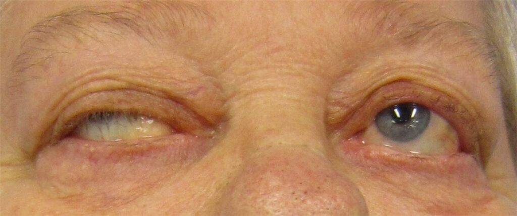
Ptosis is the presenting complaint in most cases, and its behaviour is more diagnostic than its degree. It is variable through the day, characteristically worse with fatigue and toward evening, and improved by rest or sleep.
Diplopia, when present, follows a pattern that does not fit any single cranial nerve palsy and that can shift from day to day, or even within the same examination. Bright light and sustained visual effort tend to make both worse.
On examination, ask the patient to sustain upgaze for 30 to 60 seconds and watch the ptosis worsen; this fatigue test is one of the more reliable bedside findings. Cogan’s lid twitch, a brief upward overshoot of the lid as the patient refixates from downgaze back to primary position, is specific when present but is not seen in every case.
When one lid droops significantly, the fellow lid can show compensatory retraction, a consequence of equal innervation being sent to both levator muscles under Hering’s law, and this can make an asymmetric presentation look more dramatic on the unaffected side than it actually is.
Orbicularis oculi weakness is often demonstrable as poor resistance to gentle lid opening after forced eye closure. Extraocular muscle weakness, when present, tends to follow a variable, non-anatomic pattern rather than the distribution of a single cranial nerve.
Pupils remain normal throughout, which is one of the more useful things to check at the bedside: normal pupils in a patient with ptosis and ophthalmoplegia make a compressive third nerve palsy considerably less likely.
All-fit Slit-Lamp Adapter
Record and share exactly what you see at the slit lamp. One adapter fits any slit lamp or surgical microscope — and any smartphone.
From Choroida — the team behind this siteDiagnostic Evaluation
The ice pack test is simple, well tolerated, and reasonably sensitive for myasthenic ptosis: ice applied to a ptotic lid for two minutes improves the ptosis, since neuromuscular transmission improves at lower temperature. A period of rest with the eyes closed can produce a similar improvement.
Acetylcholine receptor antibody testing is positive in most generalised disease but in a lower proportion of purely ocular presentations, which limits how much weight a negative result should carry in an ocular-only patient. MuSK antibody testing is worth checking when acetylcholine receptor antibodies are negative, particularly if there is prominent bulbar or facial involvement.
Single-fibre electromyography is the most sensitive test available and is particularly useful when serology is negative but the clinical picture, especially a positive ice pack test, remains convincing. Repetitive nerve stimulation showing a decremental response supports the diagnosis but is less sensitive than single-fibre EMG.
CT or MRI of the chest to look for thymoma is recommended in every confirmed case, ocular or generalised.
Seronegative ocular myasthenia is common enough that a negative antibody panel should not be used on its own to rule out the diagnosis when the clinical picture is otherwise convincing.
Differential Diagnosis
- Third nerve palsy, typically pupil-involving, with a fixed rather than variable pattern of restriction
- Chronic progressive external ophthalmoplegia, slowly progressive and symmetric, non-fatigable, often mitochondrial in origin, with ptosis that does not vary through the day
- Thyroid eye disease, where lid retraction rather than ptosis is the more typical finding, with restrictive rather than fatigable motility limitation
- Horner syndrome, with mild, non-fatigable ptosis and associated miosis
- Aponeurotic (involutional) ptosis, stable and non-fatigable, usually with a high eyelid crease
- Botulism and Lambert-Eaton myasthenic syndrome, other neuromuscular junction disorders with distinguishing systemic features
Variability over time is the feature most of these mimics lack. A pattern of ptosis or motility restriction that stays exactly the same from visit to visit argues against myasthenia regardless of which muscles are involved.
Management
Pyridostigmine, an acetylcholinesterase inhibitor, is typically first-line and improves neuromuscular transmission, though ocular symptoms often respond less completely to it than limb weakness does.
Prism correction or occlusion can help with residual diplopia once disease activity is otherwise controlled, though the angle’s tendency to fluctuate during active disease makes precise prism prescribing difficult until things settle.
Corticosteroids are commonly used when ocular symptoms are not adequately controlled by pyridostigmine alone, and observational data have associated steroid use with lower rates of progression to generalised disease, though whether early immunosuppression genuinely prevents generalisation or simply treats the ocular symptoms more effectively is still debated.
Steroid-sparing agents such as azathioprine or mycophenolate are used for long-term control when steroid side effects become a limiting factor.
Thymectomy is indicated for confirmed thymoma regardless of disease severity, and is considered in selected non-thymomatous generalised disease based on trial evidence. Its role in purely ocular myasthenia gravis is less well established than its role in generalised disease.
Prognosis
Patients who stay purely ocular for about two years are unlikely to generalise afterward, which is worth telling patients directly once that milestone has passed.
Ocular symptoms often respond only partially to pyridostigmine and frequently need immunosuppression for adequate control. Most patients achieve reasonable functional control of ptosis and diplopia with treatment, although relapse and fluctuation over subsequent years are common.
When generalised disease does develop, the long-term prognosis with modern immunosuppressive treatment is good, but it moves the patient’s care into closer, ongoing collaboration with neurology.


Document what you see
Two smartphone imaging tools built for everyday clinic use — one for the slit lamp, one for the fundus.
From Choroida — the team behind this siteReferences
- Kupersmith MJ. Ocular myasthenia gravis: treatment successes and failures in patients with long-term follow-up. Journal of Neurology. 2009.
- Wong SH, Huda S, Vincent A, Plant GT. Ocular myasthenia gravis: controversies and updates. Current Neurology and Neuroscience Reports. 2014.
- Nair AG, Patil-Chhablani P, Venkatramani DV, Gandhi RA. Ocular myasthenia gravis: a review. Indian Journal of Ophthalmology. 2014.
- Ocular Myasthenia Gravis. EyeWiki, American Academy of Ophthalmology.
- Myasthenia Gravis. StatPearls, NCBI Bookshelf.
Test yourself
A few questions straight from this article.
-
In ocular myasthenia gravis, disease involvement is confined to which structures?
Ocular myasthenia gravis is disease limited to the levator palpebrae and extraocular muscles. Once bulbar, limb or respiratory muscles are involved, the disease is classified as generalised. -
In most patients with ocular myasthenia gravis, autoantibodies are directed against which target?
Most patients carry antibodies to the postsynaptic acetylcholine receptor, reducing the pool of functional receptors. A smaller subset instead have muscle-specific kinase or LRP4 antibodies. -
Why are the levator palpebrae and extraocular muscles especially vulnerable in myasthenia gravis?
These muscles sustain lid position and steady fixation, firing at unusually high tonic rates. That workload, rather than any special immunological targeting of the orbit, explains why ocular signs appear first. -
Patients with ocular myasthenia gravis who remain ocular-only beyond which interval are unlikely to generalise later?
Generalisation occurs most commonly within the first two years after onset. Patients still confined to ocular disease past that milestone are statistically unlikely to generalise, and can be told so directly. -
In myasthenic ptosis, retraction of the fellow eyelid beside a drooping lid is explained by which mechanism?
Hering's law sends equal innervation to both levator muscles, so the effort to lift a heavily ptotic lid retracts the fellow lid. This makes an asymmetric case look more dramatic on the unaffected side. -
Why does cooling a ptotic eyelid improve the ptosis of ocular myasthenia gravis?
Ice applied to the ptotic lid for two minutes improves the droop because neuromuscular transmission works better at lower temperature. Simple rest with the eyes closed can produce a similar improvement. -
Which investigation is the most sensitive test available for ocular myasthenia gravis?
Single-fibre EMG is the most sensitive test and is especially valuable when serology is negative but the clinical picture remains convincing. Repetitive nerve stimulation supports the diagnosis but is less sensitive. -
Which investigation is recommended in every confirmed case of myasthenia gravis, ocular or generalised?
Chest imaging for thymoma is recommended in every confirmed case, whatever the extent of muscle involvement. A confirmed thymoma is itself an indication for thymectomy regardless of disease severity. -
In a patient with ptosis and ophthalmoplegia, which bedside finding makes a compressive third nerve palsy considerably less likely?
Pupils stay normal throughout in myasthenia gravis, whereas a compressive third nerve palsy is typically pupil-involving. Checking the pupil is one of the most useful bedside discriminators. -
Which drug is typically first-line for ocular myasthenia gravis?
Pyridostigmine, an acetylcholinesterase inhibitor, is first-line and improves neuromuscular transmission. Ocular symptoms often respond less completely than limb weakness, so corticosteroids are frequently added.

Writes and reviews the clinical articles on Educate. Every post is checked against peer-reviewed sources before it goes live.


