
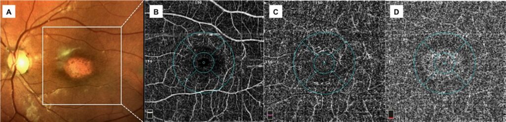
For sixty years, seeing the retinal blood vessels in detail meant injecting a dye into a patient’s arm and photographing it as it passed through the eye.
Fluorescein angiography is invasive, time-consuming, and carries a small but real risk of allergic reaction.
OCT angiography (OCTA) produces comparable, often more detailed, vascular images from a scan lasting a few seconds – with no dye, no injection, and no risk.
It does this by detecting movement rather than light absorption, which is a genuinely different way of imaging blood flow.
That difference in physics is also exactly why OCTA cannot fully replace dye angiography – each technique reveals something the other cannot.
What Is OCT Angiography?
OCT angiography is a non-invasive imaging technique that visualises retinal and choroidal blood flow by detecting motion contrast between repeated OCT scans of the same retinal location.
Understanding what it visualises requires knowing the four capillary layers it can independently image:
- The superficial capillary plexus, in the nerve fibre and ganglion cell layers
- The deep capillary plexus, at the inner nuclear layer
- The choriocapillaris, immediately beneath Bruch membrane
- In disease, an additional pathological layer such as a choroidal neovascular membrane can be segmented and visualised in isolation
This depth-resolved capability is OCTA’s defining advantage – dye angiography compresses all of these layers into a single flattened image, while OCTA can separate them.
How OCT Angiography Works
- Repeated OCT B-scans are acquired at the same retinal location in rapid succession
- Static tissue produces an identical signal on each repeated scan; moving erythrocytes within blood vessels produce a decorrelation signal between scans
- Software algorithms extract this motion contrast and generate a map of perfused vasculature, with no dye required
- Automated or manual segmentation assigns the flow signal to specific anatomical layers, producing separate en-face angiograms for each plexus
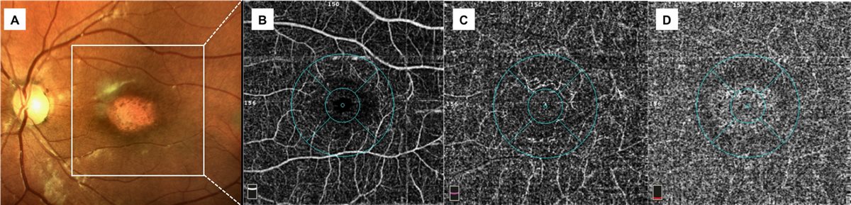
Because the whole process relies purely on detecting motion, it can be repeated as often as needed without any cumulative patient risk – a genuinely different economics of imaging compared with dye-based studies.
Fundus Explorer Pro
Photograph the retinal findings described here with the phone already in your pocket — 22 D optics and built-in illumination in one handheld unit.
From Choroida — the team behind this siteClinical Applications
Neovascular Age-Related Macular Degeneration
- Direct visualisation of choroidal neovascular membranes without waiting for dye leakage to develop, allowing faster diagnosis
- Detection of subclinical or non-exudative neovascularisation before it becomes clinically active, relevant to monitoring eyes at risk of conversion
Diabetic Retinopathy
- Visualisation of capillary non-perfusion and enlargement of the foveal avascular zone, both markers of ischaemic severity
- Detection of early microvascular changes that may precede clinically visible retinopathy on fundus examination
Vascular Occlusive Disease
- Mapping the extent of capillary non-perfusion in retinal vein and artery occlusion, informing prognosis and the risk of neovascular complications
Glaucoma
- Quantification of peripapillary and macular capillary density, an area of active research as a potential structural biomarker alongside optic nerve head OCT
The diabetic and occlusive disease applications are where OCTA has moved furthest from being a novelty toward genuine day-to-day clinical utility, because non-perfusion maps directly inform management decisions.
OCTA Versus Fluorescein Angiography
Advantages of OCTA
- Non-invasive, with no injection, no allergic reaction risk and no contraindication in renal impairment or pregnancy
- Rapid acquisition, typically a few seconds per scan, compared with several minutes for a full dye angiogram sequence
- Depth-resolved imaging of individual capillary plexuses, which dye angiography cannot separate
- Can be repeated freely for serial monitoring without cumulative risk
Limitations Compared With Dye Angiography
- Cannot show dynamic leakage, which remains the defining feature of active exudation on fluorescein angiography
- Smaller field of view in most standard acquisitions, though widefield OCTA systems are narrowing this gap
- Prone to motion, blink and segmentation artefacts that can mimic or obscure true pathology
- Cannot detect leakage from the retinal pigment epithelium or systemic sources of dye pooling in the way indocyanine green angiography can
The two techniques answer different questions – OCTA shows where vessels are and are not, while dye angiography shows where they are actively leaking – which is why most retina services use them as complementary tools rather than choosing one over the other.
Interpretation Pitfalls
- Segmentation errors, where the software misassigns flow signal to the wrong layer, are common in eyes with significant retinal structural distortion
- Projection artefact, where superficial vessel shadows appear falsely in deeper segmented layers, can mimic pathological neovascularisation
- Poor fixation, media opacity and blink artefacts degrade image quality and can produce false areas of apparent non-perfusion
- Correlating OCTA findings with the structural OCT B-scan and, where needed, with clinical examination remains essential rather than optional
Projection artefact is probably the single most common source of a false-positive neovascular membrane read on OCTA, and recognising it is a core skill for anyone interpreting these scans.
Prognosis for the Technology
OCT angiography has moved from a research tool to routine clinical equipment in a little over a decade.
- Widefield OCTA systems are progressively closing the field-of-view gap with dye angiography
- Quantitative metrics – vessel density, non-perfusion area, foveal avascular zone size – are increasingly used as trial endpoints and are being explored as early biomarkers across diabetic, occlusive and neurodegenerative disease
- Artificial intelligence-assisted artefact detection and automated quantification are active areas of development, aiming to reduce the interpretation pitfalls above
- OCTA has not eliminated the need for dye angiography, and is unlikely to for as long as dynamic leakage remains clinically important information
The realistic trajectory is complementary coexistence rather than replacement – OCTA has taken over the questions it answers better, and dye angiography has kept the ones it still answers alone.


Document what you see
Two smartphone imaging tools built for everyday clinic use — one for the slit lamp, one for the fundus.
From Choroida — the team behind this siteReferences
- Spaide RF, Fujimoto JG, Waheed NK, et al. Optical coherence tomography angiography. Progress in Retinal and Eye Research. 2018.
- Jia Y, Bailey ST, Wilson DJ, et al. Quantitative optical coherence tomography angiography of choroidal neovascularization in age-related macular degeneration. Ophthalmology. 2014.
- de Carlo TE, Romano A, Waheed NK, Duker JS. A review of optical coherence tomography angiography (OCTA). International Journal of Retina and Vitreous. 2015.
- OCT Angiography. EyeWiki, American Academy of Ophthalmology.
- Optical Coherence Tomography Angiography. StatPearls, NCBI Bookshelf.
Test yourself
A few questions straight from this article.
-
What signal does OCT angiography detect to map perfused blood vessels?
Static tissue gives an identical signal on repeated B-scans, while moving erythrocytes produce a decorrelation signal that software converts into a map of perfused vasculature. -
Which capability of OCT angiography does fluorescein angiography lack?
Depth-resolved imaging is OCTA's defining advantage: dye angiography compresses the vascular layers into a single flattened image, while OCTA can separate them. -
At which retinal layer does OCT angiography segment the deep capillary plexus?
The superficial capillary plexus lies in the nerve fibre and ganglion cell layers, while the deep capillary plexus is segmented at the inner nuclear layer. -
Which finding can OCT angiography not demonstrate, keeping fluorescein angiography in use?
OCTA shows where vessels are and are not, but cannot show dynamic leakage, which remains the defining feature of active exudation on fluorescein angiography. -
Which OCT angiography findings mark ischaemic severity in diabetic retinopathy?
OCTA visualises capillary non-perfusion and enlargement of the foveal avascular zone, both markers of ischaemic severity, and may show changes before clinically visible retinopathy. -
What advantage does OCT angiography offer in eyes at risk of converting to neovascular AMD?
OCTA can detect subclinical or non-exudative neovascularisation before it becomes clinically active, without waiting for dye leakage to develop. -
Which artefact most commonly produces a false-positive neovascular membrane on OCT angiography?
Projection artefact, where superficial vessel shadows appear falsely in deeper segmented layers, can mimic pathological neovascularisation and is the commonest false-positive source. -
How is OCT angiography currently being investigated in glaucoma?
Quantification of peripapillary and macular capillary density is an active research area as a potential structural biomarker alongside optic nerve head OCT. -
In which patients is OCT angiography free of the contraindications that apply to dye angiography?
Because no injection is given, OCTA carries no allergic reaction risk and no contraindication in renal impairment or pregnancy, and can be repeated freely. -
What is the realistic future relationship between OCT angiography and dye angiography?
OCTA has taken over the questions it answers better, but dye angiography is retained for as long as dynamic leakage remains clinically important information.

Writes and reviews the clinical articles on Educate. Every post is checked against peer-reviewed sources before it goes live.


