
A swollen, red eyelid can mean two very different things — one treatable with oral antibiotics at home, the other a genuine ophthalmic emergency.
Preseptal cellulitis is infection confined to the soft tissue in front of the orbital septum, and it is the more common — and far less dangerous — of the two.
Its twin, orbital cellulitis, involves the orbit itself and carries real risk of vision loss and intracranial spread.
The two conditions can look deceptively similar at first glance, especially through a swollen, half-closed eyelid.
What separates them is not the degree of eyelid swelling but a handful of specific findings that only a careful exam reveals.
Getting this distinction right the first time avoids both an unnecessary hospital admission and a dangerously missed emergency.
What Is Preseptal Cellulitis?
Preseptal cellulitis is infection and inflammation of the eyelid and periorbital soft tissue anterior to the orbital septum, the fibrous barrier that separates the eyelid from the orbit proper.
Its defining features include:
- Swelling, tenderness, and erythema confined to the eyelid and surrounding skin
- A white, quiet globe once the swollen lid is opened for examination
- Normal visual acuity, pupil reactivity, and eye movements
- No pain with eye movement, unlike orbital cellulitis
Because the orbital septum acts as a physical barrier, preseptal disease does not, by definition, involve the orbital contents behind it.
Epidemiology
Preseptal cellulitis is considerably more common than orbital cellulitis and carries a much lower complication rate.
- It occurs across all ages but is particularly common in young children
- Skin breaches — minor trauma, insect bites, or an infected chalazion or hordeolum — are relatively more common triggers than in orbital disease
- It can also arise from spread of adjacent infection, including sinusitis, though this is a more classic trigger for orbital rather than preseptal disease
Because most cases are mild and respond to oral therapy, many patients are managed successfully without ever being hospitalized.
All-fit Slit-Lamp Adapter
Record and share exactly what you see at the slit lamp. One adapter fits any slit lamp or surgical microscope — and any smartphone.
From Choroida — the team behind this sitePathophysiology
Infection reaches the preseptal space through a limited number of routes.
- Direct inoculation through broken skin — a cut, insect bite, or minor trauma
- Local spread from an adjacent infected structure, such as a hordeolum, dacryocystitis, or chalazion
- Hematogenous spread, now uncommon since the introduction of Haemophilus influenzae type b vaccination
- Contiguous spread from sinusitis, though this route more often produces orbital rather than purely preseptal disease
The orbital septum is the anatomical reason this infection typically stays confined to the eyelid rather than progressing to threaten the globe or optic nerve directly.
Risk Factors
Local Risk Factors
- Minor eyelid or periorbital skin trauma, including insect bites
- An untreated hordeolum, chalazion, or dacryocystitis acting as a local source
- Recent eyelid or periocular surgery
Patient-Related Risk Factors
- Young age, particularly in preschool-aged children
- Incomplete vaccination status, historically a significant risk factor before widespread Hib vaccination
- Immunosuppression, which can allow more rapid progression of an otherwise localized infection
A visible portal of entry — a scratch, insect bite, or infected eyelid lesion — is common enough in preseptal disease that its presence should be actively looked for.
Clinical Presentation
Symptoms
- Eyelid swelling, redness, and tenderness, developing over hours to a few days
- Mild fever in some patients, though high fever and systemic toxicity are more typical of orbital disease
- No pain with eye movement and no double vision
- Normal vision, even when the eyelid is too swollen to open easily
Examination Findings
- Erythematous, tender, warm eyelid swelling, often severe enough to partially or fully close the eye
- A white, non-injected globe once the lid is gently opened
- Normal visual acuity and pupil reactivity
- Full, pain-free extraocular movements
- No proptosis (forward displacement of the globe)
Every one of the “normal” findings above — vision, pupils, motility, and globe position — is what actually confirms this is preseptal rather than orbital disease.
Diagnostic Evaluation
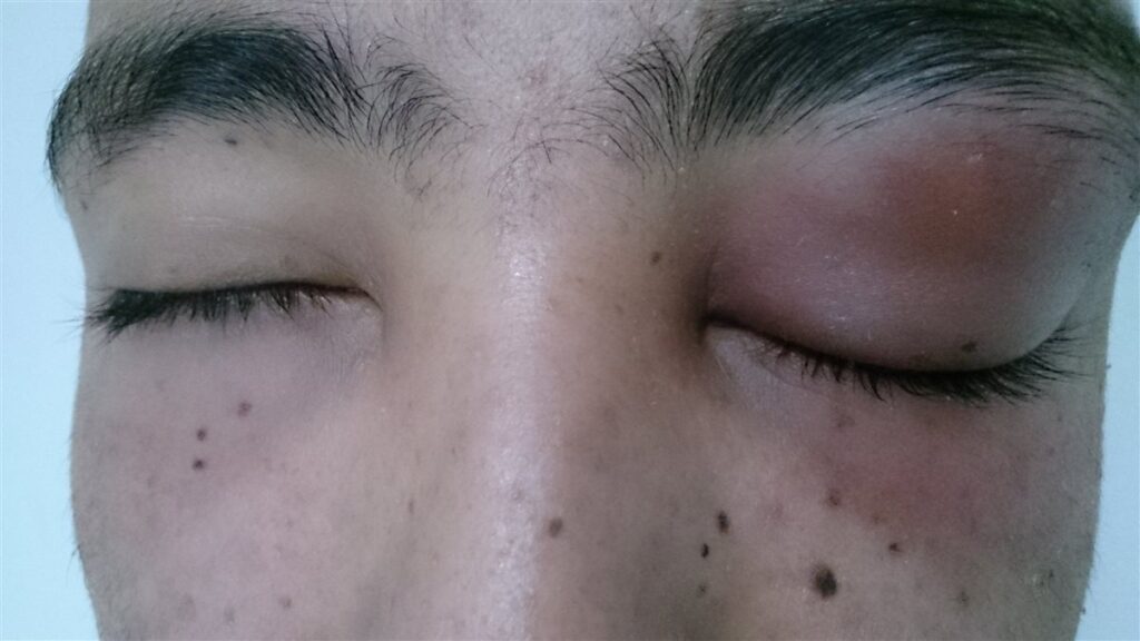
Clinical Examination
- Diagnosis is primarily clinical, based on eyelid findings combined with a genuinely normal globe examination
- Careful assessment of visual acuity, pupils, color vision, and extraocular motility in every case, however difficult the swelling makes the exam
When Imaging Is Needed
- CT of the orbits and sinuses when the exam cannot reliably exclude orbital involvement, when the child is too young or uncooperative for a full exam, or when any red-flag finding is present
- Imaging is also appropriate when the patient fails to improve on appropriate oral therapy
Laboratory Testing
- Usually not required for mild, straightforward outpatient cases
- Blood cultures have a low diagnostic yield but may be considered in more severe or systemically unwell patients
The exam, not a scan, is the primary tool for distinguishing preseptal from orbital disease — imaging is reserved for cases where the exam cannot answer the question with confidence.
Differential Diagnosis
Conditions that can be confused with preseptal cellulitis include:
- Orbital cellulitis — proptosis, restricted or painful eye movement, or reduced vision distinguishes it and represents a genuine emergency
- Allergic eyelid edema — itching predominates over pain, often bilateral, and typically not warm or markedly tender
- Hordeolum or chalazion — a discrete, localized lesion rather than diffuse eyelid swelling
- Dacryocystitis — swelling centered specifically over the lacrimal sac at the medial canthus
- Contact dermatitis — itching and a history of new topical exposure, without the acute infectious appearance
Distinguishing preseptal from orbital cellulitis is by far the most consequential differential — everything else on this list is lower stakes by comparison.
Management
Mild Disease
- Oral antibiotics covering common skin and respiratory organisms, typically amoxicillin-clavulanate, once orbital involvement has been confidently excluded
- Close outpatient follow-up, often within 24-48 hours, to confirm improvement
- Treatment of any identifiable local source, such as an infected hordeolum or wound
More Significant Disease
- Intravenous antibiotics and inpatient observation for more extensive swelling, young infants, systemic symptoms, or uncertainty about orbital involvement
- Prompt escalation to imaging and orbital cellulitis management if any red-flag finding develops during observation
Ongoing Monitoring
- Repeat assessment of vision, pupils, and motility at every follow-up, since preseptal disease can progress to orbital involvement if inadequately treated
- A low threshold to escalate care if the patient fails to improve as expected
Reassessing the eye exam at every visit — not just checking whether the swelling looks better — is what catches the uncommon case that is quietly progressing toward orbital disease.
Prognosis
Prognosis is excellent for the great majority of patients.
- Most cases resolve fully with oral antibiotics and no lasting sequelae
- Serious complications are uncommon and are essentially limited to progression to orbital cellulitis, which is a distinct and more serious diagnosis
- Vision loss and intracranial spread are risks of orbital, not preseptal, disease
The overwhelmingly favorable prognosis of preseptal cellulitis depends entirely on correctly excluding orbital involvement in the first place.
Would you like to document anterior segment findings with your smartphone?


Document what you see
Two smartphone imaging tools built for everyday clinic use — one for the slit lamp, one for the fundus.
From Choroida — the team behind this siteReferences
- Preseptal and Orbital Cellulitis. Merck Manual, Professional Edition.
- Understanding Pediatric Bacterial Preseptal and Orbital Cellulitis. Pediatrics in Review.
- Preseptal Cellulitis Versus Orbital Cellulitis: A Pediatric Case. Advanced Emergency Nursing Journal. 2022.
- Children’s Hospital of Philadelphia. Preseptal or Orbital Cellulitis Clinical Pathway.
- Preseptal Cellulitis. EyeWiki, American Academy of Ophthalmology.
Test yourself
A few questions straight from this article.
-
Preseptal cellulitis is infection confined to tissue in which location relative to the orbital septum?
The orbital septum is a fibrous barrier separating eyelid from orbit. Preseptal disease sits in front of it, which is why the orbital contents stay uninvolved. -
Which examination finding supports preseptal rather than orbital cellulitis?
It is the normal findings — vision, pupils, motility and globe position — that confirm preseptal disease. Painful or restricted movement points to orbital involvement. -
Which route of infection in preseptal cellulitis became uncommon after Hib vaccination?
Bloodborne seeding of the preseptal space is now uncommon since the introduction of Haemophilus influenzae type b vaccination. -
Which trigger is relatively more common in preseptal cellulitis than in orbital cellulitis?
Skin breaches — minor trauma, insect bites, or an infected chalazion or hordeolum — are relatively more common triggers than in orbital disease. Sinusitis is the more classic orbital trigger. -
Which oral antibiotic is typically used for mild preseptal cellulitis?
Mild disease is treated with oral antibiotics covering common skin and respiratory organisms, typically amoxicillin-clavulanate, once orbital involvement has been confidently excluded. -
How soon should a patient with mild preseptal cellulitis on oral antibiotics be reassessed?
Close outpatient follow-up, often within 24-48 hours, is used to confirm the infection is genuinely improving on oral therapy. -
When is CT of the orbits and sinuses indicated in suspected preseptal cellulitis?
Imaging is reserved for cases the exam cannot settle — an uncooperative young child, any red-flag finding, or failure to improve on appropriate oral therapy. -
Which feature distinguishes dacryocystitis from preseptal cellulitis?
Dacryocystitis produces swelling localised specifically over the lacrimal sac at the medial canthus, rather than the diffuse eyelid swelling of preseptal cellulitis. -
Which feature suggests allergic eyelid edema rather than preseptal cellulitis?
In allergic eyelid edema itching predominates over pain, the swelling is often bilateral, and the lid is typically neither warm nor markedly tender. -
Which complication accounts for essentially all serious outcomes in preseptal cellulitis?
Serious complications are uncommon and essentially limited to progression to orbital cellulitis. Vision loss and intracranial spread are risks of orbital, not preseptal, disease.

Writes and reviews the clinical articles on Educate. Every post is checked against peer-reviewed sources before it goes live.


