
DISEASE
Atrophic retinal holes are round or oval-shaped lesions often existing in the peripheral retina. They are the result of chronic atrophic changes or thinning of the sensory retina without the presence of vitreoretinal adhesions.
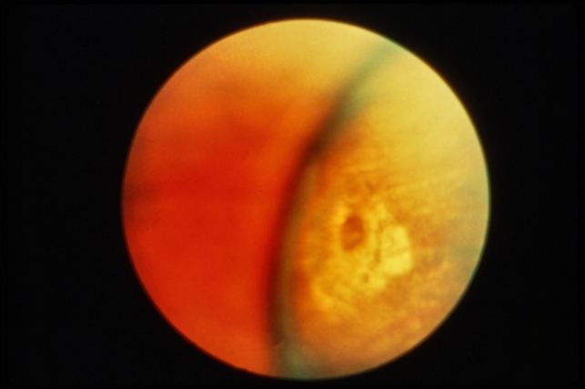
Atrophic retinal hole is a full-thickness break in the retina often present in the peripheral retina. This disease is generally associated with Lattice degeneration.
Symptoms
Most of the patients with atrophic retinal holes have no symptoms. This condition is generally found incidentally on a regular eye examination. If the patient has an associated posterior vitreous detachment, the patient may complain of decreased vision, floaters, and flashes of lights.
Fundus Explorer Pro
Photograph the retinal findings described here with the phone already in your pocket — 22 D optics and built-in illumination in one handheld unit.
From Choroida — the team behind this siteDiagnosis
Atrophic retinal holes are present on the periphery of the retina therefore the peripheral fundus examination is important in the evaluation of this disease. During the indirect ophthalmoscopic examination, the ophthalmologist put some pressure on the patient’s sclera.
This technique is called scleral indentation. Through this technique, the examiner will have a better view of the peripheral retina and the retinal holes adjacent to the ora Serrata can be easily diagnosed.
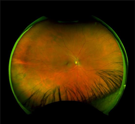
Goldmann’s three-mirror lens also provides a broader view of the peripheral fundus.
Differential Diagnosis
Atrophic retinal holes have a very typical clinical appearance of full-thickness retinal break without the presence of vitreoretinal adhesions. Despite this, there are many other diagnoses that should be considered, that include:
- Horseshoe retinal tear is full thickness, U-shaped breaks in the neurosensory retina. They occur secondary to the vitreoretinal traction.
- Lattice degeneration is a peripheral retinal degeneration characterized by localized retinal thinning.
- Operculated retinal holes are also found in the peripheral retina. They have a cap of retinal tissue pulling forward into the vitreous body where it floats above the hole.
- Snail track retinal degeneration is a group of glistening white dots found mainly on the peripheral retina.
MANAGEMENT
As these holes are small and are present in the far peripheral visual field, they are not usually even noticed or require any treatment. According to some studies, prophylactic laser retinopexy should be done in the following cases:
- if the subretinal fluid is present there.
- in myopic patients, because the chances of retinal detachment are high among these patients.
- if the retinal detachment is present in the patient’s fellow eye.


Document what you see
Two smartphone imaging tools built for everyday clinic use — one for the slit lamp, one for the fundus.
From Choroida — the team behind this siteREFERENCES
- Kanski, Jack. Clinical Ophthalmology 5th edition. Butterworth-Heinemann; 2003:359-371
- Preferred Practice Patterns: Posterior Vitreous Detachment, Retinal Breaks, and Lattice Degeneration. AAO 2008
- Byer N. Subclinical Retinal Detachment Resulting from Asymptomatic Retinal Breaks. Ophthalmology 2001; 108:1499-1504
- Gonzales C,Gupta A, Schwartz S,et al.The fellow eye of patients with phakic rhegmatogenous retinal detachment from atrophic holes of lattice degeneration without posterior vitreous detachment. Br J Ophthalmol 2004 88: 1400-1402
- Michaelson I. Role of a Distinctive Choroido-retinal Lesion in the Pathogenesis of Retinal Hole: A Clinical and Pathological Report. Br J Ophthalmol 1956 40: 527-535
- Sigelman J. Vitreous Base Classification of Retinal Tears: Clinical Application. Surv Ophthalmol 25:59-74, 1980.
Test yourself
A few questions straight from this article.
-
What process produces atrophic retinal holes?
Atrophic retinal holes result from chronic atrophic change or thinning of the sensory retina, without vitreoretinal adhesions. -
Which peripheral degeneration are atrophic retinal holes generally associated with?
An atrophic retinal hole is a full-thickness break, usually in the peripheral retina, and is generally associated with lattice degeneration. -
How are most atrophic retinal holes discovered?
Most patients with atrophic retinal holes have no symptoms, and the holes are generally found incidentally on a regular eye examination. -
When may a patient with atrophic retinal holes report floaters and flashes of light?
If there is an associated posterior vitreous detachment, the patient may complain of decreased vision, floaters and flashes of light. -
Which technique during indirect ophthalmoscopy helps detect atrophic retinal holes next to the ora serrata?
Pressing on the sclera during indirect ophthalmoscopy (scleral indentation) improves the view of the peripheral retina, so holes adjacent to the ora serrata are easily seen. -
Which lens gives a broader view of the peripheral fundus when looking for atrophic retinal holes?
Besides indirect ophthalmoscopy with indentation, the Goldmann three-mirror lens provides a broader view of the peripheral fundus. -
What distinguishes a horseshoe retinal tear from an atrophic retinal hole?
Horseshoe tears are U-shaped full-thickness breaks secondary to vitreoretinal traction, whereas atrophic holes form without vitreoretinal adhesion. -
Which feature defines an operculated retinal hole, a differential of atrophic retinal holes?
Operculated holes have a cap of retinal tissue pulled forward into the vitreous, where it floats above the hole. -
What is the usual management of atrophic retinal holes?
Because the holes are small and lie in the far peripheral field, they usually go unnoticed and do not require treatment. -
Which finding is an indication for prophylactic laser retinopexy of an atrophic retinal hole?
According to some studies, prophylactic laser retinopexy should be done when subretinal fluid is present, in myopic patients, or when the fellow eye has a retinal detachment.
