
CASE REPORT
An old man of 70 years visited an eye clinic to get a clear opinion of recently diagnosed Age-related macular degeneration (AMD). He had no significant ocular, systemic, or family history. He also said that he observed no significant difference in his vision.

His visual acuity was tested and found to be 6/9 in both of his eyes. Adnexa was marked normal. Pupils were Round, Regular, and Reactive. He presented no Metamorphopsia on the Amsler grid.
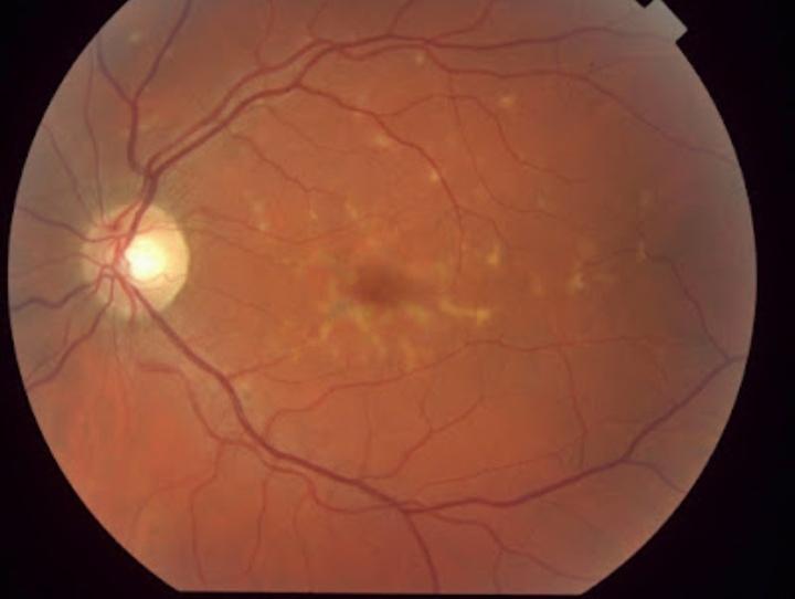
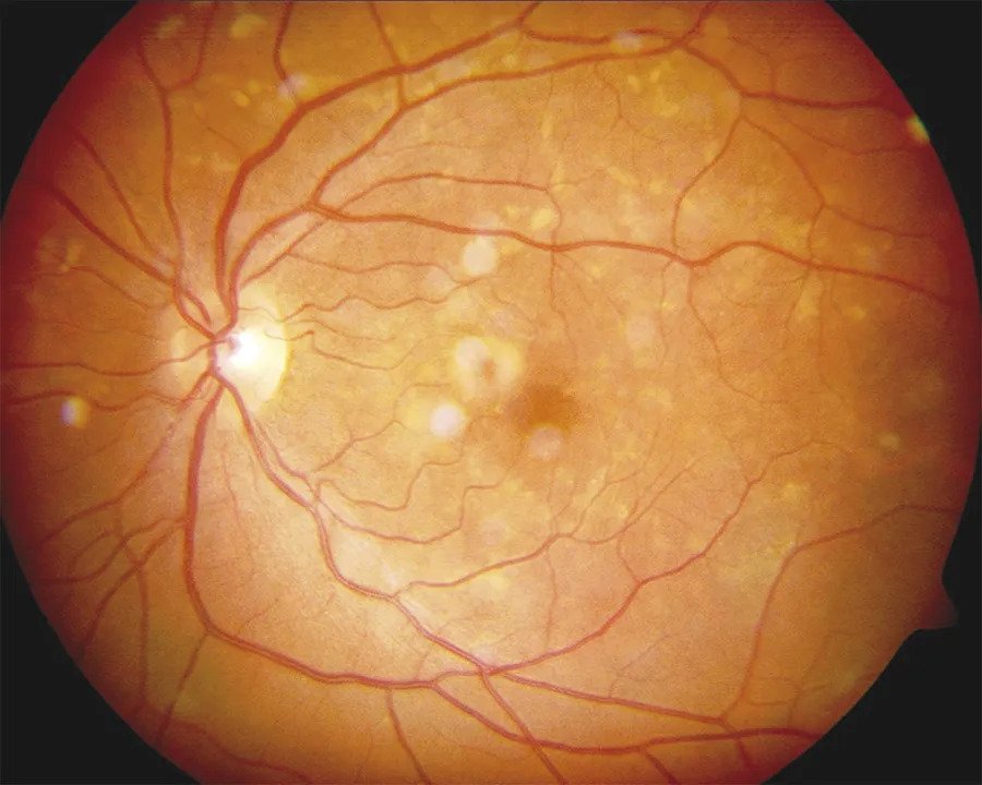
On Dilated Fundoscopy, bilateral, symmetrical sub-retinal egg yolk-like foveal lesions with drusen-like spots in the macula were seen. OCT was performed for further diagnosis. It showed bilateral hyperreflective dome-shaped elevations within RPE along with no subretinal or intra-retinal fluid and retinal edema.
On the basis of the above evaluation and examination, the patient was diagnosed with pattern dystrophy of adult-onset Foveomacular vitelliform dystrophy.
Pattern Dystrophies disease entity
Pattern dystrophies are macular diseases in which different patterns are formed due to the deposition of a lipofuscin pigment in the RPE of the macula. The onset age of these dystrophies is around 40 and 50.
These dystrophies might be misdiagnosed due to their close resemblance with age-related macular degeneration in older patients.
Pattern dystrophies occur due to mutations in the RDS gene and are considered as an autosomal dominant disease
Usually, patients are asymptomatic but with increasing age, they may show symptoms of Metamorphopsia and blurring of vision thus resembling Age-related macular degeneration.
Types of Pattern dystrophies:
Pattern dystrophies can be categorized into 5 types
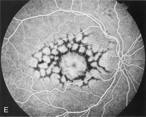
1. Reticular dystrophy:
- It is characterized by pigment deposition in the pattern of lines or knots thus giving the final shape of the fishing net.
- Initially, it affects the macula and then involves the posterior pole also.
2. Butterfly-shaped pigment dystrophy:
- It is characterized by bilateral pigment deposition in RPE in a butterfly pattern.
- These patients are mostly asymptomatic at an early age but as the age progresses these patients may face atrophic depigmented lesions extending into peripapillary regions with a marked reduction in visual acuity.
3. Adult-onset Foveomacular vitelliform dystrophy:
- It is the most common dystrophy which occurs due to faulty phagocytosis of RPE which results in the accumulation of photoreceptor debris thus leading to yellow-grey (egg yolk) macular pigmented spots and lesions of Retinal Pigment Epithelium (RPE).
4. Multifocal pattern dystrophy simulating Stargardt disease:
- It is characterized by irregular, multiple yellow-white deposits which are scattered throughout the posterior pole thus resembling the shape of flecks seen in Stargardt disease.
- Mild to moderate constriction of the peripheral visual field may be noted.
- It can be differentiated from Stargardt disease in the following aspects.
It is an autosomal dominant disease, which occurs in the late fifties and patients with this disease have good visual acuity and dark choroid is also absent.
5. Fundus pulverulentus:
- It is considered the rarest of all dystrophies.
- It is characterized by pigment mottling of RPE and pigment deposits occur in the form of granules.
Pattern Dystrophies MANAGEMENT
There is no treatment for the accumulation of lipofuscin in the various pattern dystrophies. Fortunately, vision loss is slowly progressive compared to AMD. However, vision loss progresses more rapidly in some patients than in others.
As there is no treatment to reverse vision loss, patients may benefit from low-vision aids and therapy. Similar to AMD, treatment modalities are focused primarily on complications that may arise from RPE degeneration with the most common yet infrequent one being CNV.
CNV arising from pattern dystrophies has been shown to be responsive to anti-VEGF therapy. Treatment with anti-VEGF therapy has been shown to decrease macular edema and improve visual acuity.
Treatment of CNV with PDT should be considered with caution as vitilleform lesions treated with PDT without any components of CNV may have a negative impact on vision.
Adult vitelliform dystrophy patients can develop macular holes due to the degeneration of overlying outer retinal and photoreceptor layers. Treatment with vitrectomy has mostly been ineffective.
Would you have interest in taking retina images by smartphone?
Fundus photography is superior to fundus analysis as it enables intraocular pathologies to be photo-captured and encrypted information to be shared with colleagues and patients.
Recent technologies allow smartphone-based attachments and integrated lens adaptors to transform the smartphone into a portable fundus camera and Retinal imaging by smartphone.
RETINAL IMAGING BY YOUR SMARTPHONE
REFERENCES
- Sjogren H. Dystrophia Reticularis Laminae Pigmentosae Retinae. Acta Ophthalmol. 1950;28:279-295.
- Zhang K, Garibaldi DC, Li Y, et al. Butterfly-Shaped Pattern Dystrophy: A Genetic, Clinical, and Histopathololgical report. Ophthlmic Mol Genet. 2002;120:485-490
- Gass JDM. A clinicopathologic study of a peculiar foveomacular dystrophy. Trans Am Ophthalmol Soc 1974; 72: 139–156.
- Deutman, A. F., van Blommestein, J. D. A., Henkes, H. E., Waardenburg, P. J., Solleveld-van Driest, E. Butterfly-shaped pigment dystrophy of the fovea. Arch. Ophthal. 83: 558-569, 1970.
- Gass JMD. Stereoscopic Atlas Of Macular Disease. Philadelphia, Pa:Elsvier; 1997.
- Kim RY, Dollfus H, Keen TJ, et al. Autosomal Dominant Pattern Dystrophy of the Retina Associated with a 4-basepair insertion at Codon 140 in the Peripherin/RDS gene. Arch Ophthalmol. 113, 451-455.
