
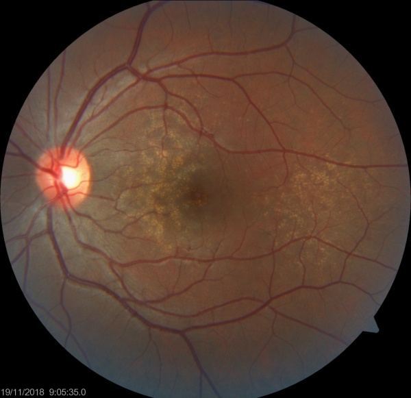
CASE REPORT OF BASAL LAMINAR DRUSEN
A 65-year-old woman complained of blurred vision in both eyes. At her first ophthalmic examination, her visual acuity was 20/20 in both eyes. Ophthalmoscopy showed many small sub–RPE deposits in both eyes.
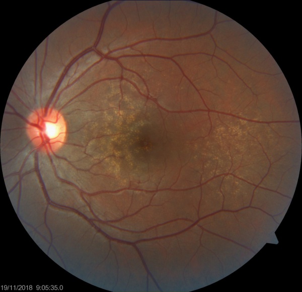
The SD-OCT images showed RPE nodular elevations which had a ‘saw-tooth pattern. This blunted triangular or prolapsed shape has been reported to be typical for cuticular drusen.
Reticular pseudodrusen were also seen in both eyes. The scotopic and photopic full-field electroretinograms were reduced, and Goldmann perimetry showed a decrease in the sensitivity in the pericentral field in both eyes. she was diagnosed with cuticular drusen, and placed on a periodic follow-up examination schedule.
DISEASE “BASAL LAMINAR DRUSEN”
Basal laminar drusen/Cuticular drusen is an uncommon entity. Although originally thought to be distinct from age-related macular degeneration, it is now considered to be in the same spectrum as macular degeneration.
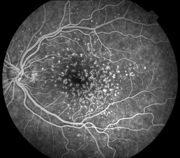
Findings include numerous small yellow drusen on examination, which are more obvious on fundus auto-fluorescence. Fluorescein angiography (FA) demonstrates punctuate areas of early staining throughout the macula, with a “stars in the sky” pattern.
Vitelliform macular lesions development, choroidal neovascularization (CNV), and geographic atrophy (GA) may cause vision loss.
Fundus Explorer Pro
Photograph the retinal findings described here with the phone already in your pocket — 22 D optics and built-in illumination in one handheld unit.
From Choroida — the team behind this siteDIAGNOSIS
Basal laminar drusen diagnosis is clinical, based on dilated fundus examination, fundus autofluorescence, and FA.
MANAGEMENT OF BASAL LAMINAR DRUSEN
Patients with basal laminar drusen should be monitored for the development of “pseudovitelliform” retinal pigment epithelium lesions/detachment, CNV, or GA which may be visually significant.
The complete pathophysiology of BLD has not been elucidated and a CFH mutation has been implicated in the development of membranoproliferative glomerulonephritis type 2 (MPGN type II) (dense deposit disease) and atypical hemolytic uremic syndrome (aHUS).
It is recommended that renal function be monitored in those presenting with BLD, especially at an early age (between 5 and 30 years).
In addition, glomerulonephritis with isolated C3 deposits, a condition that is an intermediate between MPGN and aHUS, also has an association with CFH mutation.
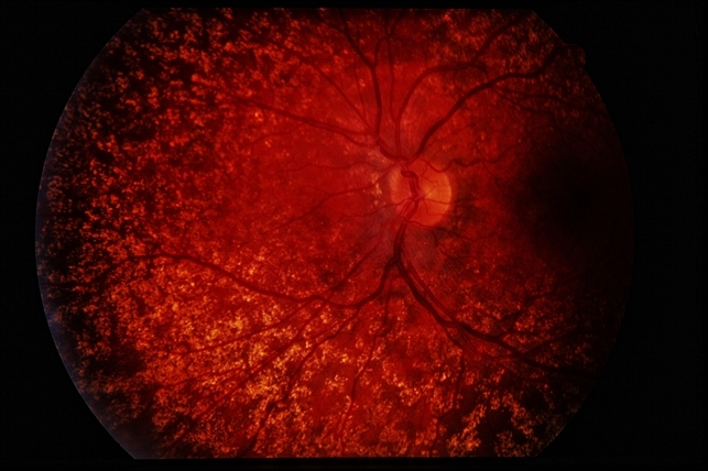
There does not appear to be a correlation between the severity of disease within the kidney and the eye, though 80% of patients with dense deposit disease present with cuticular drusen.
Similarly, genetic analysis for certain mutations may be beneficial to help guide certain patients with known alleles associated with disease progression.


Document what you see
Two smartphone imaging tools built for everyday clinic use — one for the slit lamp, one for the fundus.
From Choroida — the team behind this siteREFERENCES
- 1. Boon, C. J., van de Kar, N. C., Klevering, B. J., Keunen, J. E., Cremers, F. P., Klaver, C. C., Hoyng, C. B., Daha, M. R., & den Hollander, A. I. (2009). The spectrum of phenotypes caused by variants in the CFH gene. Molecular Immunology, 46(8–9), 1573–1594.
- Russell SR, Mullins RF, Schneider BL, Hageman GS. (2000). Location, substructure, and composition of basal laminar drusen compared with drusen associated with aging and age-related macular degeneration. American Journal of Ophthalmology, 129:205-14.
- Grassi MA, Folk JC, Scheetz TE, Taylor CM, Sheffield VC, Stone EM. (2007). Complement factor H polymorphism p.Tyr402His and cuticular Drusen. Arch Ophthalmology, 125:93-7.
- Barbazetto, I. A., Yannuzzi, N. A., Klais, C. M., Merriam, J. E., Zernant, J., Peiretti, E., Yannuzzi, L. A., & Allikmets, R. (2007). Pseudo-Vitelliform Macular Detachment and Cuticular Drusen: Exclusion of 6 Candidate Genes. Ophthalmic Genetics, 28(4), 192–197.
- Balaratnasingam, C, Cherepanoff, S., Dolz-Marco, R., Killingsworth, M.,Chen, F.K., Mendis, R., Mrejen, S. Too, L.K., Gal-Or, O., Curcio, C.A., Freund, K.B., & Yannuzzi, L.A. (2018). Cuticular drusen: Clinical phenotypes and natural history defined using multimodal imaging. Ophthalmology, 125, 100–118.
Test yourself
A few questions straight from this article.
-
How are basal laminar (cuticular) drusen currently classified in relation to age-related macular degeneration?
Although basal laminar drusen were originally thought to be a distinct entity, they are now regarded as lying within the same spectrum as macular degeneration. -
Which fundus appearance is characteristic of basal laminar drusen?
Examination shows numerous small yellow drusen, and these become far more obvious on fundus autofluorescence than on colour photography alone. -
What pattern does fluorescein angiography show in basal laminar drusen?
Fluorescein angiography demonstrates punctate areas of early staining scattered throughout the macula, described as a 'stars in the sky' pattern. -
Which SD-OCT appearance is reported as typical of cuticular drusen?
SD-OCT shows nodular elevations of the retinal pigment epithelium in a saw-tooth pattern, a blunted triangular or prolapsed shape reported as typical for cuticular drusen. -
On what is the diagnosis of basal laminar drusen based?
The diagnosis is clinical, resting on dilated fundus examination together with fundus autofluorescence and fluorescein angiography. -
Which developments should patients with basal laminar drusen be monitored for?
Vision loss in this condition comes from pseudovitelliform pigment epithelial lesions or detachment, choroidal neovascularization, and geographic atrophy, so follow-up targets those three. -
A mutation in which gene links basal laminar drusen to systemic renal disease?
A complement factor H mutation has been implicated in membranoproliferative glomerulonephritis type 2 (dense deposit disease) and in atypical haemolytic uraemic syndrome. -
Which additional monitoring is recommended for patients presenting with basal laminar drusen, especially at ages 5 to 30?
Because of the complement factor H association with glomerulonephritis and atypical haemolytic uraemic syndrome, renal function should be monitored, particularly when the drusen appear between 5 and 30 years of age. -
What proportion of patients with dense deposit disease present with cuticular drusen?
Eighty per cent of patients with dense deposit disease present with cuticular drusen, even though the severity of the renal and ocular disease does not correlate. -
What role does genetic analysis play in patients with basal laminar drusen?
Testing for certain mutations may be beneficial in guiding management for patients carrying known alleles associated with disease progression.
