Macular edema after retinal detachment surgery is a common reason for poor vision when the retina has reattached.
It appears in a minority of eyes and often follows scleral buckling or vitrectomy with gas or oil.
OCT makes the diagnosis easy, but the cause and the treatment need careful thought.
How Common It Is
Early studies found cystoid macular edema in a small share of eyes after scleral buckling, and later series reported rates up to 10 percent or more.
Lobes and Grand described the incidence after buckling in the 1980s.
Meredith and colleagues reported similar findings after detachment repair.
Modern OCT detects subclinical edema more often than older angiography (see cystoid macular edema).
Causes of Macular Edema After Retinal Detachment Surgery
Several mechanisms may act together.
- Inflammation after surgery, cryotherapy, and laser
- Disruption of the blood-retinal barrier
- Epiretinal membrane or vitreomacular traction
- Silicone oil and other tamponade agents
Sabates and colleagues described macular changes after detachment surgery, including edema and pucker.
Fundus Explorer Pro
Photograph the retinal findings described here with the phone already in your pocket — 22 D optics and built-in illumination in one handheld unit.
From Choroida — the team behind this siteRisk Factors for Macular Edema After Retinal Detachment Surgery
Risk rises with macula-off detachment, long duration of detachment, and preoperative PVR.
Surgery with extensive cryotherapy and repeated procedures also increases the risk.
Combined cataract surgery may add to inflammation, so the edema may have mixed causes (see Irvine-Gass syndrome).
Diagnosis of Macular Edema After Retinal Detachment Surgery
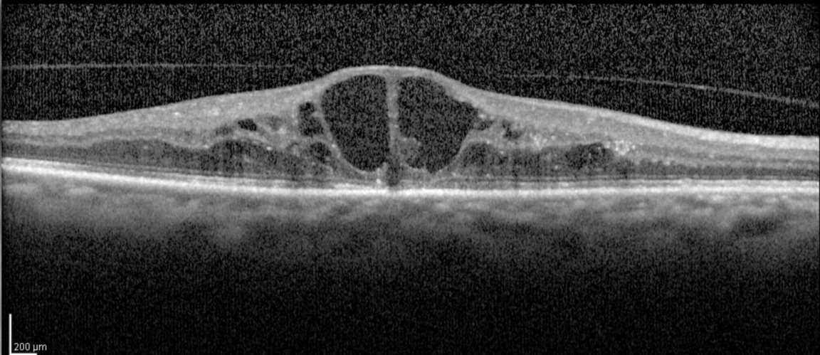
OCT shows cystic spaces in the outer plexiform and inner nuclear layers, with retinal thickening.
Look for an epiretinal membrane, a residual subretinal fluid pocket, or traction.
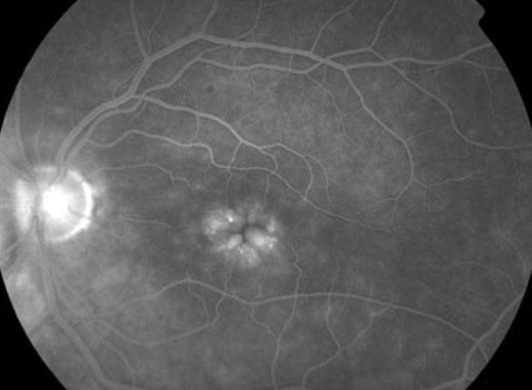
Fluorescein angiography shows petaloid leakage, and it helps when the cause is unclear.
Check the retina for a new break or redetachment, since fluid may signal failure.
Treatment of Macular Edema After Retinal Detachment Surgery
Many cases improve over time, so observation is reasonable for mild edema.
Topical steroids and nonsteroidal drops are the first treatment, and they may be combined.
Persistent edema may need a periocular or intravitreal steroid, or an anti-VEGF injection.
Peel an epiretinal membrane when traction explains the edema (see epiretinal membrane).
Prognosis of Macular Edema After Retinal Detachment Surgery
Many eyes regain vision after the edema resolves, though some develop outer retinal damage.
Persistent edema after months carries a worse outlook.
Monitor with OCT every few weeks until the macula stabilizes.
An Example Case
A 56-year-old man undergoes vitrectomy and gas for a macula-off retinal detachment, and the retina stays attached.
At three months, his acuity is 20/60, and OCT shows cystic edema with a small epiretinal membrane.
He starts topical steroid and nonsteroidal drops, and the edema improves partly after six weeks.
Because a membrane remains, the surgeon peels it, and the edema resolves, with 20/40 vision.
Questions Patients Ask
Patients ask why vision is blurry when the retina is attached, and the answer is that fluid collects in the macula.
They also ask whether the swelling will go away, and many cases improve over months.
Because some patients worry about another operation, explain that drops and injections come first.
Pitfalls to Avoid
- Assuming that every blurred vision after surgery is slow recovery
- Skipping OCT when acuity fails to improve
- Starting steroids without checking pressure and infection
- Overlooking a membrane that needs peeling
Looking Ahead
Better imaging and new drugs may help to target the cause of edema.
Also, trials may clarify whether prophylactic drops reduce the risk after surgery.
Until then, treat by cause and monitor closely.
Practical Points
Obtain OCT at the first sign of poor recovery after surgery.
Then check for epiretinal membrane, subretinal fluid, and traction.
Also review the drop regimen and the intraocular pressure at each visit.
Differential Diagnosis in Brief
Residual subretinal fluid, a macular hole, and redetachment may all reduce vision after surgery.
OCT separates them from cystic edema.
Therefore, always examine the retina and the macula together.
Preventing Edema After Detachment Surgery
Use gentle cryotherapy and laser.
Also treat inflammation early with topical steroid drops.
Because membranes can contribute, remove them during primary surgery when possible.
Then follow the patient with OCT in the first weeks.
Counselling
Explain that vision may improve slowly over several months.
Also tell patients to report any new distortion or loss of vision.
Because edema may recur after tapering drops, taper slowly.
When to Operate
Consider surgery when OCT shows a persistent membrane with traction and the vision falls.
Also consider it when medical treatment fails after three months.
Because the procedure carries risks, discuss them with the patient.
For the latest studies, see the PubMed results for macular edema after retinal detachment surgery.


Document what you see
Two smartphone imaging tools built for everyday clinic use — one for the slit lamp, one for the fundus.
From Choroida — the team behind this siteReferences
- Lobes LA Jr, Grand MG. Incidence of cystoid macular edema following scleral buckling surgery. Arch Ophthalmol. 1980;98:1230-1232.
- Meredith TA, Reeser FH, Topping TM, Aaberg TM. Cystoid macular edema after retinal detachment surgery. Ophthalmology. 1980;87:1090-1095.
- Sabates NR, Sabates FN, Sabates R, et al. Macular changes after retinal detachment surgery. Am J Ophthalmol. 1989;108:22-29.
- American Academy of Ophthalmology. Basic and Clinical Science Course, Section 12: Retina and Vitreous. AAO; current edition.

Writes and reviews the clinical articles on Educate. Every post is checked against peer-reviewed sources before it goes live.