Welding arc maculopathy is a photic injury of the fovea after viewing an electric arc.
The arc emits intense ultraviolet and blue light.
Therefore, unprotected viewing can damage the central retina within seconds to minutes.
Source of the Injury
Arc welding produces a plasma with very bright light across the spectrum.
Short wavelengths cause photochemical damage in photoreceptors and the pigment epithelium.
Also, heat adds a minor part.
As a result, the injury is mainly photochemical.
Who Gets Welding Arc Maculopathy
Welders and nearby workers are the main victims.
Bystanders and apprentices also suffer when they look at the arc.
Also, hobbyists who weld without a proper shield are at risk.
Therefore, ask about exposure in unexplained central loss.
Fundus Explorer Pro
Photograph the retinal findings described here with the phone already in your pocket — 22 D optics and built-in illumination in one handheld unit.
From Choroida — the team behind this siteMechanism of Welding Arc Maculopathy
Blue light excites chromophores such as lipofuscin and rhodopsin.
They generate reactive oxygen species that damage the outer retina.
Also, the fovea receives the full focus of the light.
As a result, the damage localizes to the center.
Photosensitizing Drugs
Some drugs raise sensitivity to light.
Power and colleagues described welding arc maculopathy with fluphenazine.
Also, phenothiazines and psoralens may play a role.
Therefore, review the medication list in every patient.
Symptoms of Welding Arc Maculopathy
Patients report a central blur, a small scotoma or a color change in the center.
Symptoms begin within hours of the exposure.
Also, pain is usually absent in the retinal form.
In contrast, arc eye causes pain from corneal injury.
Welding Flash and the Cornea
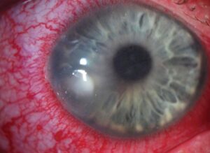
Ultraviolet light injures the corneal epithelium and causes photokeratitis.
Pain, tearing and photophobia start hours later.
Also, the retina may be injured at the same time.
Therefore, examine the fundus in all welders with flash injury.
Fundus Findings
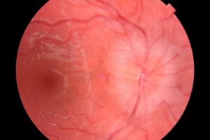
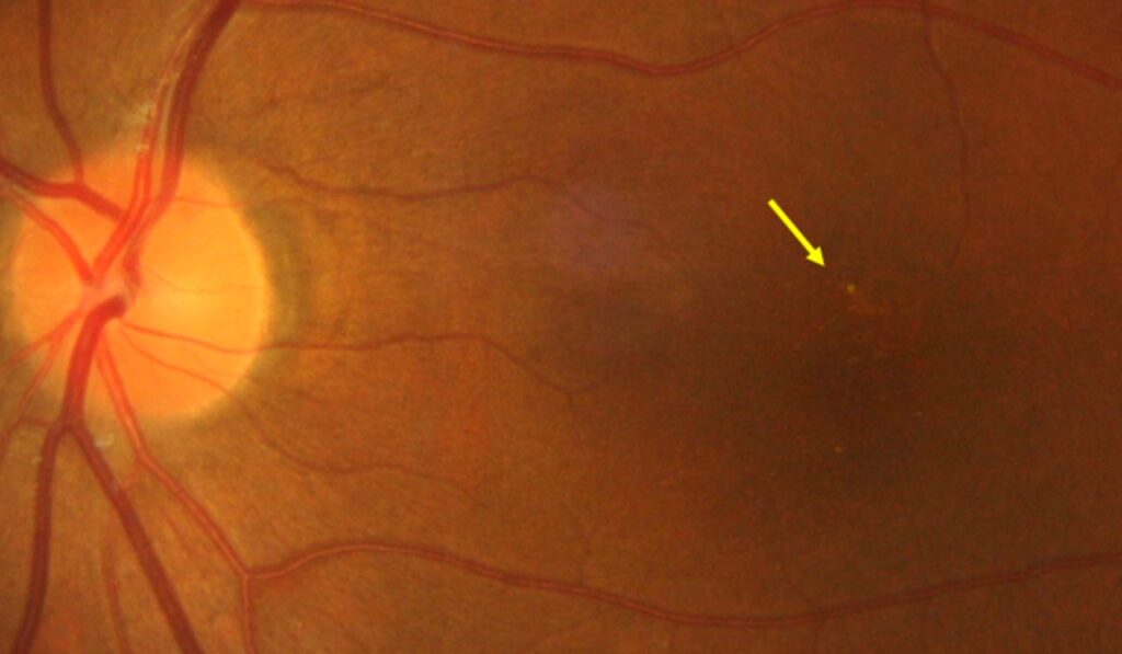
Early signs include a small yellowish or gray spot at the fovea.
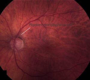
The lesion may fade and leave a pigment change or a lamellar defect.
Also, the fundus can look almost normal in mild cases.
As a result, OCT is a key test.
OCT Findings of Welding Arc Maculopathy
OCT shows disruption of the ellipsoid zone and the outer retina at the fovea.
Hyperreflective changes may appear in the outer nuclear layer.
Also, the retinal thickness can decrease later.
Therefore, scans track recovery (see solar retinopathy).
Comparison with Solar Retinopathy
Both conditions involve photochemical damage to the fovea.
Solar retinopathy follows sun gazing, and the lesion is often smaller and more symmetrical.
Also, Yannuzzi and colleagues described the solar pattern in detail.
In addition, welding injury may include a wider area.
Differential Diagnosis
Laser pointer injury and other photic injuries give similar images.
Central serous disease and macular holes also mimic them.
Also, a clear history of exposure points to the diagnosis.
Therefore, combine history with OCT.
Natural Course of Welding Arc Maculopathy
Many patients recover most of their vision over weeks to months.
Final acuity often lies between 20/20 and 20/40.
Also, a small paracentral scotoma may remain.
As a result, reassure patients while following them.
Treatment
No treatment has proven benefit.
Steroids and antioxidants have been tried without clear evidence.
Also, observation is the standard.
In addition, advise protection from further exposure.
Preventing Welding Arc Maculopathy
Use a welding helmet with a filter of the correct shade for arc work.
Auto-darkening filters help when the arc starts.
Also, bystanders need shields or screens.
Therefore, workplace rules protect the eyes.
Protective Standards
Filter shades between 10 and 14 are used for most arc welding.
The right shade depends on the current and process.
Also, check that the helmet has no cracks or light leaks.
As a result, regular inspection is part of safety.
Counseling
Explain the cause and the usual recovery.
Warn that repeated exposures raise the risk of lasting damage.
Also, advise a follow-up visit and a repeat scan.
Therefore, patients understand both the risk and the outlook.
Pitfalls to Avoid
- Do not skip the fundus examination in a welder with flash pain
- Never forget to ask about photosensitizing drugs
- Do not promise full recovery
- Never rely on sunglasses as arc protection
Documentation and Workplace Reports
Record the date, the duration of exposure and the shield used.
Report the injury to the employer or safety officer as local rules require.
Also, workers’ compensation may apply.
Therefore, good records protect the patient.
Visual Field and Amsler Testing
Use an Amsler grid or microperimetry to map the central defect.
Repeat at each visit to track recovery.
Also, document the size of the scotoma.
In addition, check color vision in the center.
Return to Work
Allow patients to return to work only with proper eye protection.
Advise a short rest from welding while symptoms last.
Also, review the helmet and the work setup.
Therefore, prevention is part of care.
For the latest studies, see the PubMed results for welding arc maculopathy.


Document what you see
Two smartphone imaging tools built for everyday clinic use — one for the slit lamp, one for the fundus.
From Choroida — the team behind this siteReferences
- Naidoff MA, Sliney DH. Retinal injury from a welding arc. Am J Ophthalmol. 1974;77:663-668.
- Power WJ, Travers SP, Mooney DJ. Welding arc maculopathy and fluphenazine. Br J Ophthalmol. 1991;75:433-435.
- Yannuzzi LA, Fisher YL, Krueger A, Slakter J. Solar retinopathy: a photobiologic and geophysical analysis. Trans Am Ophthalmol Soc. 1987;85:120-158.
- Youssef PN, Sheibani N, Albert DM. Retinal light toxicity. Eye (Lond). 2011;25:1-14.

Writes and reviews the clinical articles on Educate. Every post is checked against peer-reviewed sources before it goes live.