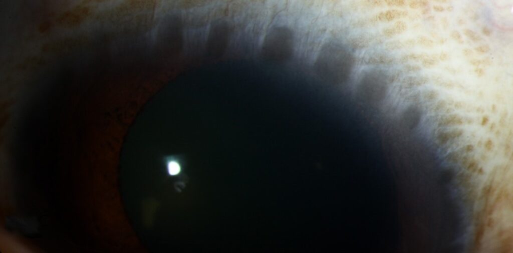
Herbert’s pits are small, shallow depressions at the upper limbus that remain after the limbal follicles of trachoma heal.
They are among the few signs that prove past trachoma on their own.
Therefore, a careful look at the limbus is worth the extra minute in any patient from an endemic area.
What Herbert’s Pits Are
Active trachoma produces follicles on the upper tarsal conjunctiva and sometimes at the superior limbus.
Those limbal follicles carry the name Herbert’s follicles.
When a follicle resolves, it leaves a small pit with a rim of scarred tissue.
Because the epithelium and the superficial stroma heal with scar, the pit stays for life.
How to Find Herbert’s Pits
Ask the patient to look down and lift the upper lid gently.
Then inspect the superior limbus under slit-lamp magnification with a narrow beam.
Pits show as tiny clear or slightly pigmented dimples along the limbal edge.
Also, some patients have a band of superficial vessels, called pannus, extending above the pits.
Finally, evert the lid, because Herbert’s pits usually coexist with Arlt’s line and diffuse tarsal scarring.
All-fit Slit-Lamp Adapter
Record and share exactly what you see at the slit lamp. One adapter fits any slit lamp or surgical microscope — and any smartphone.
From Choroida — the team behind this siteHerbert’s Pits in the Differential Diagnosis
Other conditions can leave marks at the limbus, so keep the list short.
Vernal keratoconjunctivitis produces Horner-Trantas dots, which are transient white clumps of eosinophils, which differ from pits (see limbal vernal keratoconjunctivitis).
Phlyctenulosis leaves a triangular scar after a small nodule heals (see phlyctenular keratoconjunctivitis).
Terrien marginal degeneration produces a peripheral gutter, which differs from discrete pits.
What Herbert’s Pits Mean for Management
Herbert’s pits show past disease but not active infection.
Active infection shows as follicles and inflammation, so look for TF and TI before you treat.
Because scarring that creates pits may also distort the lid, check the lashes for trichiasis (see trichiasis).
In endemic areas, screen household members, since children carry the reservoir of infection.
Documenting the Limbus
Record how many pits you see and where they sit.
Most appear at the upper limbus, between the ten and two o’clock positions.
Also, note any pannus above them, since pannus reflects chronic inflammation.
A slit-lamp photograph keeps a permanent record for follow-up.
Linking the Limbus to the Lid
Pits rarely occur alone.
When you find them, evert the upper lid and look for follicles or scars.
Also, check the lashes for trichiasis, since scarring and pits come from the same process.
Because the chain from follicles to scars to trichiasis takes years, early detection gives you time to intervene.
Telling Pits From Dellen
Dellen are shallow areas of thinning next to an elevated limbal lesion.
They stain with fluorescein and resolve after lubrication (see corneal dellen).
Pits, in contrast, stay unchanged and keep their shape.
Therefore, review the patient after a week of lubricants if you doubt the diagnosis.
Why the Sign Matters in the Field
Because the pits persist for life, they give historical evidence in survey work.
Programmes still base decisions on active signs, such as the prevalence of TF in children, when they plan mass azithromycin treatment.
Even so, a trained examiner who sees pits in an adult knows that the community had trachoma years ago.
Practical Summary for the Clinician
Look for pits at the upper limbus, then evert the lid.
Grade the lid with the WHO system and note any trichiasis.
Also, treat only active disease, and arrange surgery when lashes touch the globe.
Pannus and Corneal Changes
Chronic trachoma leads to superficial vascularization of the upper cornea, called pannus.
A fibrovascular pannus can reach the pupil and reduce vision.
Pits sit at the limbus, where the pannus begins.
Because pannus progresses in untreated disease, it indicates how long the inflammation lasted.
Natural History of Limbal Follicles
Limbal follicles appear in active trachoma, usually in the upper limbus.
They begin as small gelatinous nodules, and then they flatten and scar.
Therefore, a patient with pits has had disease long enough for follicles to form and heal.
History Questions Worth Asking
Ask where the patient grew up, about childhood eye disease, and about family members with in-turned lashes.
Also, ask about past eyelid surgery, because surgery signals advanced scarring.
Limits of the Sign
Not every patient with healed trachoma has pits, so absence does not exclude the diagnosis.
Also, a young child from an endemic area may have active disease without pits.
Therefore, always grade the lid.
For the latest studies, see the PubMed results for Herbert’s pits.


Document what you see
Two smartphone imaging tools built for everyday clinic use — one for the slit lamp, one for the fundus.
From Choroida — the team behind this siteReferences
- Taylor HR, Burton MJ, Haddad D, West S, Wright H. Trachoma. Lancet. 2014;384:2142-2152.
- Thylefors B, Dawson CR, Jones BR, West SK, Taylor HR. A simple system for the assessment of trachoma and its complications. Bull World Health Organ. 1987;65:477-483.
- Dawson CR, Jones BR, Tarizzo ML. Guide to Trachoma Control in Programmes of Primary Health Care. World Health Organization; 1981.
- Solomon AW, Zondervan M, Kuper H, Buchan JC, Mabey DCW, Foster A. Trachoma Control: A Guide for Programme Managers. World Health Organization; 2006.

Writes and reviews the clinical articles on Educate. Every post is checked against peer-reviewed sources before it goes live.