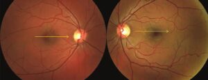
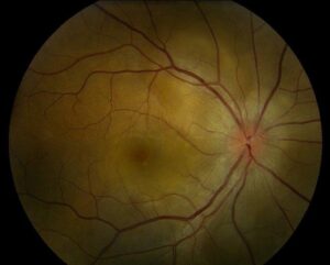
Benign concentric annular macular dystrophy is a rare dominant disorder that produces a ring of depigmentation around the fovea.
Because vision stays relatively good for many years, Deutman called it benign.
The bull’s-eye appearance raises concern for drug toxicity, so the history and tests are important.
What Benign Concentric Annular Macular Dystrophy Is
Deutman described the disorder in 1974 in a family with an annular macular lesion and mild symptoms.
Also, the disease appears in the third to fifth decade, with gradual decline in vision.
Therefore, many patients retain acuity around 20/30 to 20/60 for years (see pattern dystrophies).
Clinical Features of Benign Concentric Annular Macular Dystrophy
- A concentric annular zone of RPE atrophy around a spared fovea
- Mild metamorphopsia and a paracentral ring scotoma
- Normal peripheral retina
- Dominant family history in most cases
Gass illustrated the lesion in his atlas, and Kanski summarizes its appearance (see central areolar choroidal dystrophy).
Fundus Explorer Pro
Photograph the retinal findings described here with the phone already in your pocket — 22 D optics and built-in illumination in one handheld unit.
From Choroida — the team behind this siteInvestigations in Benign Concentric Annular Macular Dystrophy
First, fluorescein angiography shows a ring-shaped window defect.
Also, autofluorescence shows a ring of reduced signal with a normal center.
Then OCT shows outer retinal thinning in the annulus with preserved fovea in early disease.
ERG is usually normal, though multifocal ERG may show a ring of loss.
Genetics of Benign Concentric Annular Macular Dystrophy
Many families carry mutations in PRPH2, the gene for peripherin 2.
Michaelides and colleagues reviewed the genetics of inherited macular dystrophies, including this group.
The same gene causes a broad range of phenotypes, so variable expression occurs within families.
Differential Diagnosis of Benign Concentric Annular Macular Dystrophy
- Hydroxychloroquine and chloroquine toxicity
- Stargardt disease and cone dystrophy
- Central areolar choroidal dystrophy
- Occult macular dystrophy
Take a careful drug history, and test the family when needed (see hydroxychloroquine toxicity).
Management of Benign Concentric Annular Macular Dystrophy
Because no treatment exists, care focuses on diagnosis and counseling.
Also provide low vision support as needed.
Then offer genetic counseling to families with a dominant pattern.
An Example Case
A 46-year-old woman reports mild blurring and a faint ring in her central vision.
Examination shows an annular depigmented zone around a spared fovea in both eyes, with acuity of 20/40.
She takes no hydroxychloroquine or other toxic drugs, and her mother has a similar appearance.
Electrophysiology shows mildly reduced macular function, and genetic testing finds a pathogenic PRPH2 variant.
Questions Patients Ask
Patients ask whether they will go blind, and the disease usually progresses slowly with preserved central vision for years.
They also ask whether the ring will grow, and some patients show slow enlargement.
Because the name says benign, explain that some patients still lose vision later.
Pitfalls to Avoid
- Calling every bull’s-eye maculopathy toxic without history
- Missing a dominant family pattern
- Skipping a drug review for chloroquine and related agents
- Overlooking Stargardt disease and cone dystrophy as alternatives
Looking Ahead
Better imaging and genetic panels will clarify how this entity fits within PRPH2-related disease.
Also, trials of gene therapy for PRPH2 may emerge.
Therefore, precise diagnosis becomes more valuable.
Practical Points
Photograph the macula, and obtain OCT and autofluorescence.
Then confirm with genetic testing when possible.
Also follow the patient every year to document change.
Distinguishing From Toxic Maculopathy
Toxic ring maculopathy follows drug exposure and often shows parafoveal loss on OCT.
Dominant dystrophy runs in families and starts without drug use.
Because both can look alike, history is the key.
Visual Function Tests
Visual fields show a paracentral ring scotoma.
Multifocal ERG shows a ring of reduced response.
Therefore, functional tests support the diagnosis.
Following the Patient Over Time
Examine the patient yearly with OCT and autofluorescence.
Because the ring may expand slowly, measure its diameter.
Also repeat visual fields every one to two years.
Then record any change in acuity or symptoms.
Role of Electrophysiology
Full-field ERG usually remains normal, which separates this disease from cone and cone-rod dystrophies.
However, multifocal ERG shows a ring of reduced response that matches the lesion.
Therefore, use electrophysiology to confirm the diagnosis and to exclude generalized disease.
Counselling the Family
Explain that the course is usually mild.
Also tell relatives that testing is available.
Because anxiety is common, give written information.
Summary
A bull’s-eye ring in a young patient without drug exposure suggests this dystrophy.
Then confirm with imaging and genetics.
Because the course is mild, reassure while documenting change.
Also review drug history at each visit.


Document what you see
Two smartphone imaging tools built for everyday clinic use — one for the slit lamp, one for the fundus.
From Choroida — the team behind this siteReferences
- Deutman AF. Benign concentric annular macular dystrophy. Am J Ophthalmol. 1974;78:384-396.
- Michaelides M, Hunt DM, Moore AT. The genetics of inherited macular dystrophies. J Med Genet. 2003;40:641-650.
- Gass JDM. Stereoscopic Atlas of Macular Diseases: Diagnosis and Treatment. 4th ed. Mosby; 1997.
- American Academy of Ophthalmology. Basic and Clinical Science Course, Section 12: Retina and Vitreous. AAO; current edition.

Writes and reviews the clinical articles on Educate. Every post is checked against peer-reviewed sources before it goes live.