Oxalosis retinopathy is the retinal deposition of calcium oxalate crystals in patients with primary hyperoxaluria or severe kidney failure.
It produces refractile crystals, pigment changes, and macular scarring, and it often signals advanced systemic disease.
Recognition helps in diagnosis and in managing the dangerous multisystem disease behind it.
What Oxalosis Retinopathy Is
Primary hyperoxaluria is a group of rare genetic disorders of glyoxylate metabolism, and it leads to overproduction of oxalate.
The kidney fails to clear the excess, and calcium oxalate crystals deposit in kidneys, bones, heart, and eyes.
Cochat and Rumsby reviewed its types and the importance of early diagnosis (see crystalline deposits in cystinosis).
Secondary oxalosis can occur with kidney failure and high oxalate intake.
Fundus Findings in Oxalosis Retinopathy
Meredith and colleagues described the retinal signs in children and adults with the disease.
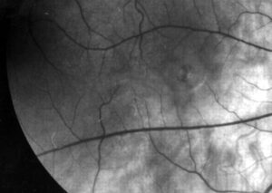
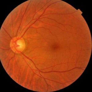
- Small, refractile white crystals in the posterior pole, often near vessels
- Retinal pigment epithelium hyperplasia, with a bull’s-eye pattern in some eyes
- Macular scarring, with pigment clumps
- Optic atrophy or disc swelling in severe cases
The crystals may be difficult to see in early disease, so good illumination helps.
Fundus Explorer Pro
Photograph the retinal findings described here with the phone already in your pocket — 22 D optics and built-in illumination in one handheld unit.
From Choroida — the team behind this siteImaging
OCT shows hyperreflective deposits in the inner and outer retina, and disruption of the RPE.
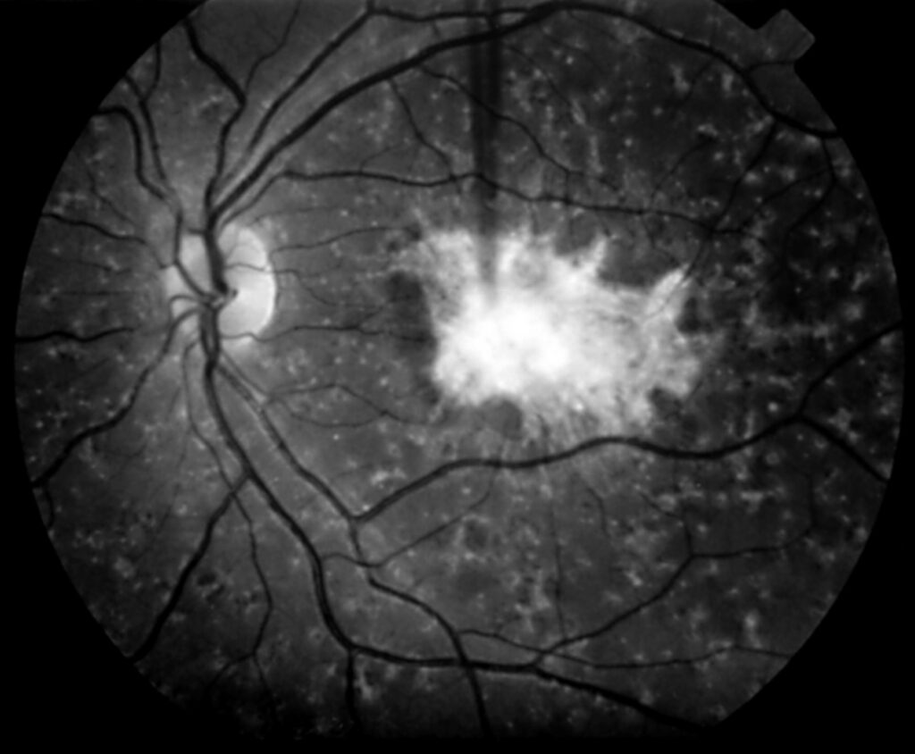
Autofluorescence reveals RPE damage, with hypoautofluorescent areas where atrophy has developed.
Fluorescein angiography may show window defects from RPE loss (see fundus autofluorescence imaging).
Differential Diagnosis
- Crystalline retinopathy from tamoxifen
- Bietti crystalline dystrophy
- Cystinosis and talc retinopathy
- Canthaxanthin retinopathy
The medical history and kidney function give the best clues (see Bietti crystalline dystrophy).
Management of Oxalosis Retinopathy
There is no specific retinal treatment, and care focuses on controlling oxalate levels.
Therefore, hydration, pyridoxine, and in some cases combined liver and kidney transplantation reduce oxalate burden.
Also, ophthalmologists should monitor the retina and the optic nerve, and coordinate with the nephrology team.
Retinal findings may regress slightly after transplantation, but scars remain.
Prognosis of Oxalosis Retinopathy
However, vision loss correlates with macular involvement and optic atrophy.
Also, early treatment of the underlying disease may preserve vision, and screening of at-risk children helps.
Explain to families that retinal changes reflect systemic disease and do not require retinal surgery.
An Example Case
A 12-year-old boy with kidney failure from primary hyperoxaluria has blurred central vision.
The fundus shows many small refractile crystals in the macula, with pigment clumping.
OCT shows hyperreflective deposits, and the family learns that the retina reflects the systemic oxalate burden.
He receives a combined liver and kidney transplant, and his retinal findings stay stable, with partial regression of the crystals.
Questions Patients Ask
Patients ask whether the crystals can be removed, and no eye treatment removes them.
They also ask whether vision will recover, and scars limit recovery.
Because the disease affects many organs, explain the role of the metabolic and kidney teams.
Pitfalls to Avoid
- Attributing crystals to drug toxicity without checking kidney function
- Missing the diagnosis in young patients with unexplained renal failure
- Delaying referral until the macula scars
- Treating the retina while ignoring oxalate control
Looking Ahead
New drugs that lower oxalate production may change the disease course.
Also, early screening of relatives may prevent advanced retinopathy.
Therefore, retinal findings may become less common in the future.
Practical Points
Document the fundus with photographs and OCT at baseline.
Then repeat them after major treatment changes.
Also share the images with the nephrology team.
Systemic Features
Patients develop kidney stones, nephrocalcinosis, and renal failure.
Also, crystals deposit in bone, heart, nerves, and vessels, so the disease needs multidisciplinary care.
Because infants may present with kidney failure, the retina may show crystals early.
Diagnostic Tests for Oxalosis Retinopathy
Measure urinary and plasma oxalate, and perform genetic testing for AGXT, GRHPR, and HOGA1 variants.
Then confirm the type, because treatment differs.
Also test relatives when a mutation is found.
Treatment of the Systemic Disease
High fluid intake, citrate, and pyridoxine help in some types.
Combined liver and kidney transplantation corrects the metabolic defect in type 1 disease.
Newer RNA interference drugs lower oxalate production.
Therefore, refer patients early, and monitor the eyes while treatment evolves.
Ophthalmic Follow-Up
Examine the eyes at diagnosis and then every six to twelve months.
Because crystals may increase with high oxalate levels, check after major treatment changes.
Also use OCT to follow macular thickness and scars.
Then share the findings with the nephrology team.
Counselling Families
Explain that the retina reflects the whole-body disease.
Because outcomes depend on early treatment, urge families to follow the medical plan.
Also discuss low vision support if the macula scars.
Finally, give the family a contact for questions.


Document what you see
Two smartphone imaging tools built for everyday clinic use — one for the slit lamp, one for the fundus.
From Choroida — the team behind this siteReferences
- Small KW, Letson R, Scheinman J. Ocular findings in primary hyperoxaluria. Arch Ophthalmol. 1990;108:89-93.
- Meredith TA, Wright JD, Gammon JA, et al. Ocular involvement in primary hyperoxaluria. Arch Ophthalmol. 1984;102:584-587.
- Cochat P, Rumsby G. Primary hyperoxaluria. N Engl J Med. 2013;369:649-658.
- American Academy of Ophthalmology. Basic and Clinical Science Course, Section 12: Retina and Vitreous. AAO; current edition.

Writes and reviews the clinical articles on Educate. Every post is checked against peer-reviewed sources before it goes live.