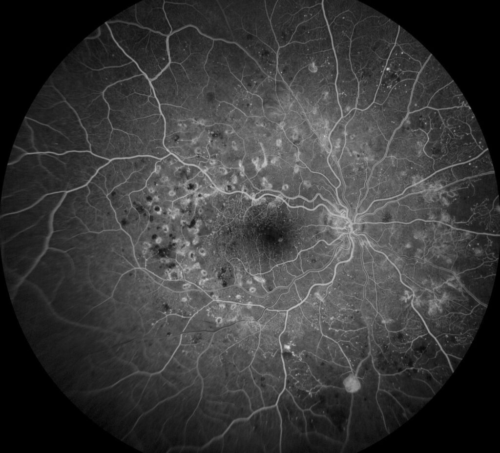
DRCR Protocol S asked whether an anti-VEGF injection could replace scatter laser for proliferative diabetic retinopathy.
It was the first large randomized trial to test this question, and its answer changed how PDR is managed.
It also carried a warning about patients lost to follow-up.
Why DRCR Protocol S Was Needed
Panretinal photocoagulation has been the standard treatment for decades, but it causes loss of peripheral field and night vision.
It can worsen macular edema (see the Diabetic Retinopathy Study).
Anti-VEGF drugs were known to make new vessels regress, so researchers tested whether doctors could use them as first-line treatment.
DRCR Protocol S: The Design
- 305 participants, 394 eyes, with previously untreated proliferative retinopathy, with or without macular edema
- Investigators randomized eyes to PRP in one to three sittings, or to intravitreal ranibizumab 0.5 milligrams, given monthly at first and then as needed
- Eyes with center-involving edema and decreased vision in the ranibizumab group received treatment for edema. Those in the PRP group received ranibizumab for the edema
- The main outcome was the change in visual acuity at two years, with a noninferiority margin of five letters (see the references)
Fundus Explorer Pro
Photograph the retinal findings described here with the phone already in your pocket — 22 D optics and built-in illumination in one handheld unit.
From Choroida — the team behind this siteVisual Acuity Results
At two years, the mean change in acuity was about 2.8 letters in the ranibizumab group and 0.2 letters in the PRP group, which met the criterion for noninferiority.
The upper bound showed that ranibizumab was not worse than laser, and it even suggested a slight advantage.
DRCR Protocol S: Other Results
- Less peripheral visual field loss with ranibizumab
- Fewer vitrectomies for hemorrhage or traction
- A lower rate of new center-involving macular edema
- Similar regression of new vessels, though some regrowth in the ranibizumab group if injections lapsed
The number of injections in the ranibizumab group was about 7 in the first year and a lower number later.
Five-Year Outcomes
At five years, visual acuity outcomes were similar between groups, and the ranibizumab group maintained its advantage in field preservation and edema (see the references).
However, investigators lost many patients to follow-up, in particular in the ranibizumab arm, and some of them had serious outcomes.
DRCR Protocol S: What Changed in Practice
- Anti-VEGF therapy became an accepted first-line option for PDR, especially in patients with macular edema or who cannot tolerate laser
- Laser remained a good choice for patients who may not attend regular visits
- Combination plans were used, with laser added after stabilization (see anti-VEGF therapy)
The Follow-Up Lesson
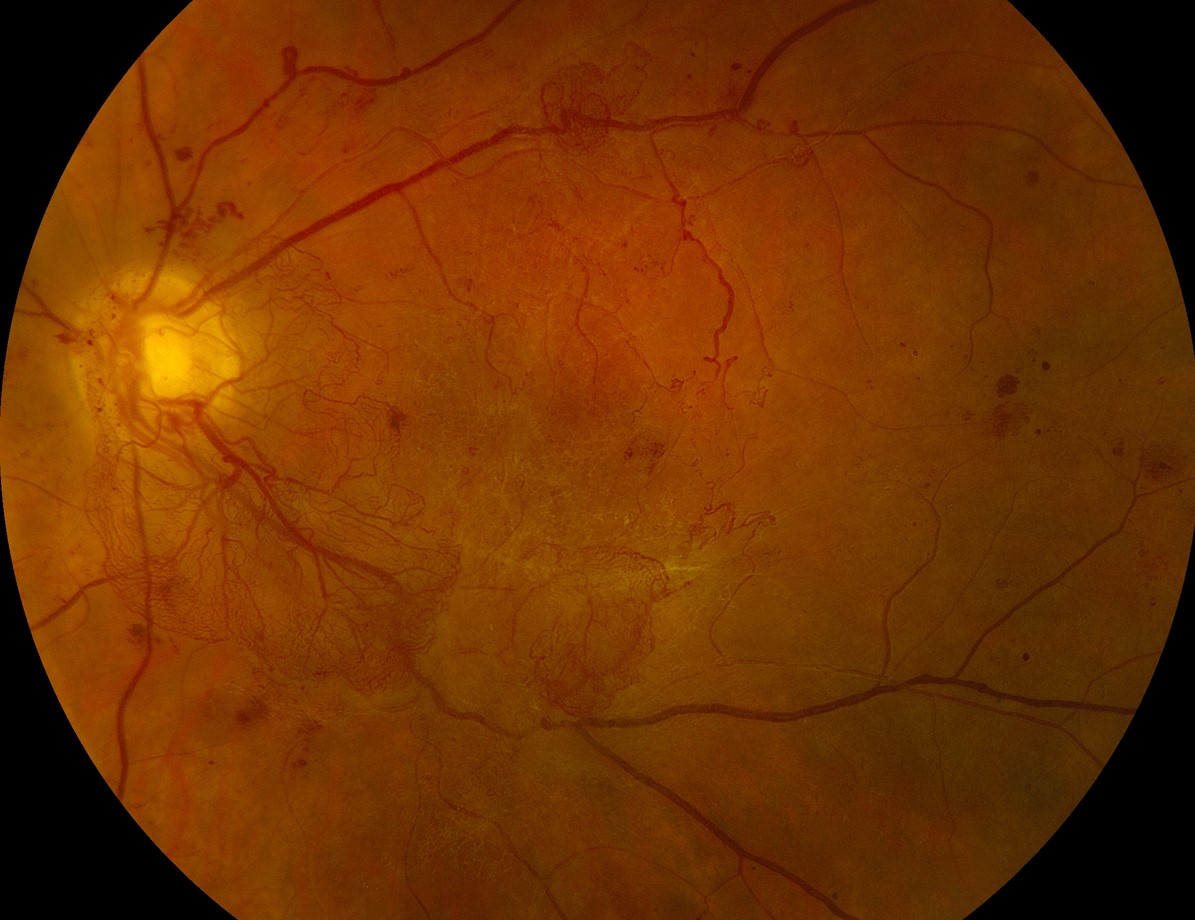
Anti-VEGF drugs are temporary, and when injections stop, new vessels can return and bleed.
Patients lost to follow-up in the trial lost vision, so the choice of treatment should consider the reliability of the patient.
DRCR Protocol S: Limits
The trial excluded patients with severe vitreous hemorrhage and traction, and the follow-up was limited to five years.
It tested ranibizumab, and results for other drugs come from other studies.
Counselling
Explain the options, including the need for repeated injections, and the fact that laser is a single, permanent treatment with side effects on the periphery.
Make a plan for follow-up, and document the discussion.
Practical Decision Making
- Prefer anti-VEGF: patients with center-involving edema, those with a need to preserve peripheral field, such as drivers and pilots. Those who can attend regular visits
- Prefer PRP: patients with poor attendance, those who cannot afford repeated injections, and pregnant women, in whom anti-VEGF drugs are used with caution
- Combine: inject first to treat edema or reduce bleeding, and add PRP to give a lasting effect (see panretinal photocoagulation)
What Happens If Injections Stop?
New vessels regress with anti-VEGF drugs but can regrow within weeks to months when injections lapse.
Protocol S patients who missed visits developed bleeding and, in some cases, tractional detachment, so a plan for reminders and a laser backup is wise.
Counselling Points
- Explain the number of visits in the first year, which is often seven or more
- Describe the risk of losing contact with the clinic, and offer to treat with laser if attendance is a concern
- Discuss pregnancy plans and other drugs
Cost and Access
The trial did not weigh cost, but it matters in practice: laser is cheaper in the long run, while injections have an upfront burden for each visit.
Documentation and Planning
Record the baseline field, the presence of edema, and the plan for follow-up, since these details guide the choice.
Review pregnancy status, which changes the options, and write down who will remind the patient of visits.
For the latest studies, see the PubMed results for DRCR Protocol S.


Document what you see
Two smartphone imaging tools built for everyday clinic use — one for the slit lamp, one for the fundus.
From Choroida — the team behind this siteReferences
- Writing Committee for the Diabetic Retinopathy Clinical Research Network; Gross JG, Glassman AR, Jampol LM, et al. Panretinal photocoagulation vs intravitreous ranibizumab for proliferative diabetic retinopathy: a randomized clinical trial. JAMA. 2015;314:2137-2146.
- Gross JG, Glassman AR, Liu D, et al. Five-year outcomes of panretinal photocoagulation vs intravitreous ranibizumab for proliferative diabetic retinopathy: a randomized clinical trial. JAMA Ophthalmol. 2018;136:1138-1148.
- Ryan SJ, Schachat AP, Wilkinson CP, et al, eds. Retina. 6th ed. Elsevier; 2018.
- American Academy of Ophthalmology. Basic and Clinical Science Course, Section 12: Retina and Vitreous. AAO; current edition.

Writes and reviews the clinical articles on Educate. Every post is checked against peer-reviewed sources before it goes live.