DRCR Protocol I is the trial that moved ranibizumab to the center of treatment for diabetic macular edema.
Before it, focal laser was the standard, and steroid injections were an alternative with side effects.
The trial compared all of them in the same study, and gave a clear ranking.
Why DRCR Protocol I Was Needed
The ETDRS showed that laser slows vision loss in macular edema, but it rarely improved vision (see the ETDRS).
Small studies suggested that anti-VEGF injections could do better, and steroids had shown mixed results (see intravitreal triamcinolone).
DRCR Protocol I: The Design
The Diabetic Retinopathy Clinical Research Network enrolled 854 eyes with center-involving edema and visual acuity of 20/32 to 20/320.
- Sham injection plus prompt laser
- Ranibizumab 0.5 milligrams plus prompt laser
- Ranibizumab plus deferred laser, starting after at least 24 weeks
- Triamcinolone 4 milligrams plus prompt laser
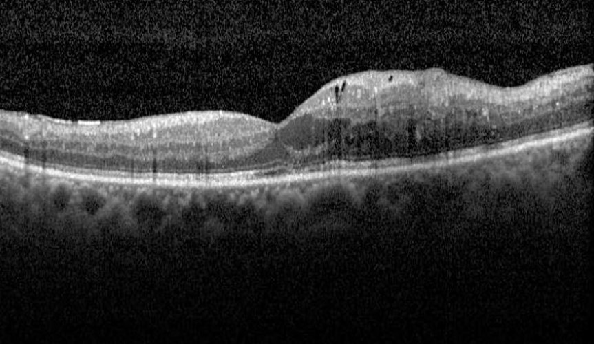
Retreatment followed a protocol based on OCT and acuity.
Fundus Explorer Pro
Photograph the retinal findings described here with the phone already in your pocket — 22 D optics and built-in illumination in one handheld unit.
From Choroida — the team behind this siteOne-Year Results
- Mean acuity change was about +9 letters in both ranibizumab groups, compared with +3 letters for sham plus laser
- Triamcinolone gave an average of about +4 letters, not better than laser overall
- In pseudophakic eyes, triamcinolone performed as well as ranibizumab, while phakic eyes had cataract and pressure rise (see the references)
DRCR Protocol I: Two- and Five-Year Results
At two years, the advantage of ranibizumab was maintained, and the deferred laser group did as well as the prompt laser group, with fewer laser treatments.
At five years, the gains were maintained in eyes that continued treatment, with fewer injections needed over time (see the references).
The study suggested that laser could be held in reserve, and that injections were the key part of treatment.
Safety
- Endophthalmitis was rare in ranibizumab groups
- Systemic adverse events were similar in groups
- Triamcinolone raised intraocular pressure and caused cataract surgery in a large share of phakic eyes
DRCR Protocol I: What Changed in Practice
- Anti-VEGF drugs replaced laser as first-line treatment for center-involving edema with reduced vision
- Laser was reserved for selected cases and as an adjunct
- Monthly injections at first, then a treat-and-extend plan, became the usual approach (see treat-and-extend regimen)
- Steroids were kept for pseudophakic eyes and for eyes that fail anti-VEGF drugs
Limits
The trial tested ranibizumab only, and head-to-head comparisons of other drugs came later, including Protocol T (see DRCR Protocol T).
Visual acuity of 20/32 or worse was required, so eyes with good vision were studied in a later trial (see DRCR Protocol V).
DRCR Protocol I: Counselling
Explain that injections are the best way to improve vision in edema, that many are needed in the first year, and that fewer are needed later.
Stress that blood sugar and blood pressure control remain essential.
Why the Result Was Surprising
Laser had been the standard for twenty years, and few expected injections to produce gains of nearly two lines in a month-long course.
The trial also showed that waiting to give laser did not harm vision, and fewer laser treatments were required in the deferred group, which suggested that the injections were doing most of the work (see anti-VEGF therapy).
How Treatment Was Delivered
- Monthly injections in the first four months or until stability
- Retreatment decisions based on OCT thickness and visual acuity at each visit
- Laser in the deferred group only after at least 24 weeks, if edema persisted
After the first year, the number of injections fell, to about three in the second year and fewer after that.
Using the Findings Today
- Begin with injections in eyes with center-involving edema and reduced vision (see diabetic macular edema)
- Consider laser when edema persists with focal leaking microaneurysms, and keep it away from the fovea
- Use a steroid when anti-VEGF drugs fail, mostly in pseudophakic eyes
Practical Cautions
- Examine the pressure after injection and watch for endophthalmitis
- Review systemic control, since high blood pressure and kidney disease worsen edema
- Check for tractional elements on OCT before giving more injections
Registry and Real-World Data
Real-world series show that many patients receive fewer injections than in the trial, and that results are often a little worse, mainly because of missed visits and delayed retreatment.
Teams that track attendance and send reminders reproduce the trial results more closely, so a recall system is part of treatment.
Retinal specialists should document the thickness and acuity at every injection, because retreatment decisions in the trial depended on these two numbers, and a clear record makes each choice easier to justify.
Patients who see steady gains by month six are likely to need fewer injections later.
For the latest studies, see the PubMed results for DRCR Protocol I.


Document what you see
Two smartphone imaging tools built for everyday clinic use — one for the slit lamp, one for the fundus.
From Choroida — the team behind this siteReferences
- Elman MJ, Aiello LP, Beck RW, et al. Randomized trial evaluating ranibizumab plus prompt or deferred laser or triamcinolone plus prompt laser for diabetic macular edema. Ophthalmology. 2010;117:1064-1077.
- Elman MJ, Qin H, Aiello LP, et al. Intravitreal ranibizumab for diabetic macular edema with prompt versus deferred laser treatment: three-year randomized trial results. Ophthalmology. 2012;119:2312-2318.
- Ryan SJ, Schachat AP, Wilkinson CP, et al, eds. Retina. 6th ed. Elsevier; 2018.
- American Academy of Ophthalmology. Basic and Clinical Science Course, Section 12: Retina and Vitreous. AAO; current edition.

Writes and reviews the clinical articles on Educate. Every post is checked against peer-reviewed sources before it goes live.