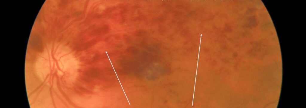
New treatments for retinal vein occlusion target the macular edema that causes most of the vision loss.
Anti-VEGF drugs became the standard after the first trials, and newer drugs extend the interval or give another route of treatment.
Also, the choice depends on the type of occlusion, the response, and the patient’s ability to attend.
New Treatments for Retinal Vein Occlusion: Standard Treatment Today
Ranibizumab and aflibercept improve vision in both branch and central vein occlusion, as shown in the BRAVO, CRUISE, COPERNICUS, and GALILEO trials (see the references and central retinal vein occlusion).
Also, bevacizumab is a lower-cost alternative, and the SCORE2 and LEAVO trials supported its use in many patients (see the references).
Laser is also an option for branch occlusion with persistent edema or ischemic areas (see branch retinal vein occlusion).
Faricimab
Faricimab, the bispecific antibody against VEGF-A and angiopoietin-2, has been tested in the BALATON and COMINO trials in branch and central occlusion.
In addition, results showed noninferior vision to aflibercept, with a strong drying effect and the potential for extended dosing intervals.
It is approved in several regions for this use.
Fundus Explorer Pro
Photograph the retinal findings described here with the phone already in your pocket — 22 D optics and built-in illumination in one handheld unit.
From Choroida — the team behind this siteNew Treatments for Retinal Vein Occlusion: High-Dose Aflibercept
Trials of aflibercept 8 milligrams in vein occlusion test whether longer intervals can maintain the benefit, building on results in AMD and diabetic edema (see high-dose aflibercept).
Approval depends on the region, and clinicians should also follow the local label.
Steroid Implants
- A dexamethasone implant lasts several months and gave benefit in the GENEVA trials, with fewer injections than anti-VEGF drugs (see dexamethasone intravitreal implant)
- It suits pseudophakic eyes, patients who cannot attend often, and eyes that respond poorly to anti-VEGF drugs
- Pressure rise and cataract are the main risks
New Treatments for Retinal Vein Occlusion: Combination and Sequential Approaches
Some eyes need anti-VEGF plus steroid to control edema, and a switch from one drug to another may recover response.
Laser may be added to treat leaking microaneurysms in branch occlusion.
Treating the Underlying Cause
- A full medical workup, including blood pressure, glucose, lipids, and clotting tests in younger patients (see retinal vein occlusion workup)
- Control of glaucoma, which raises risk
- Treatment of ischemia and neovascularization, with scatter laser and injections when needed (see neovascular glaucoma)
Choosing Among New Treatments for Retinal Vein Occlusion
- Start with an anti-VEGF drug in most eyes with center-involving edema
- Extend intervals as the retina dries, using treat-and-extend
- Add or switch to a steroid in refractory or pseudophakic eyes
- Consider longer-acting drugs for patients with limited access
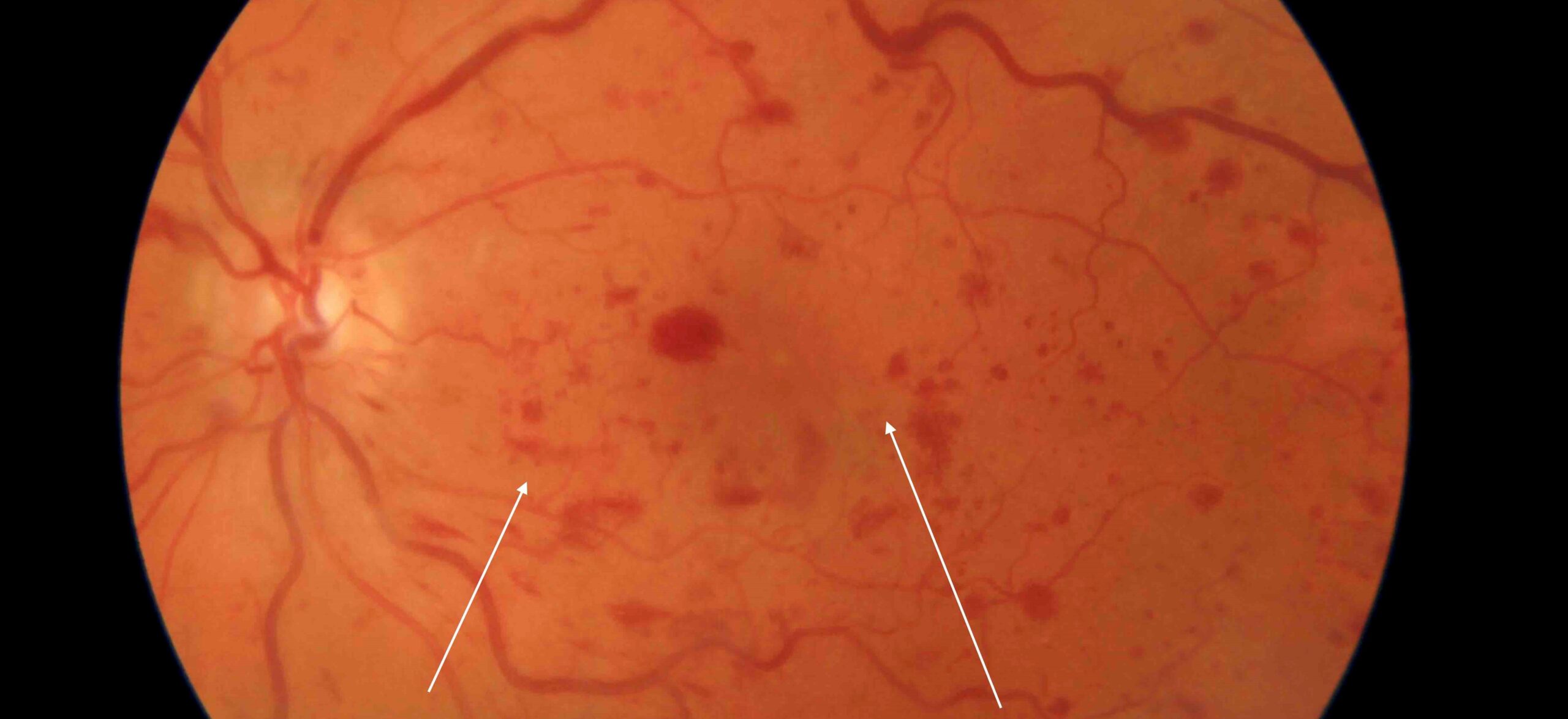
New Treatments for Retinal Vein Occlusion: Counselling
Explain that edema often needs treatment for a year or more, and that missed visits risk vision loss.
Also, advise patients that control of blood pressure and other conditions reduces the risk of another event.
Cost and Treatment Burden
Many patients with vein occlusion are older, have other illnesses, and live far from the clinic.
Treatment plans should consider travel and the cost of drugs, and use treat-and-extend or longer-acting drugs where suitable.
Bevacizumab keeps the cost down, and the LEAVO trial found that it was less effective than aflibercept for central occlusion, so the choice is a balance.
Monitoring and Retreatment
- Review monthly at first, with OCT and visual acuity
- Extend the interval only when the macula has been dry for a period
- Check for neovascularization on the iris and angle, particularly in ischemic central occlusion
- Treat ischemic areas with laser if neovascularization develops
Example Case
A 65-year-old man with central vein occlusion and 20/200 vision receives monthly aflibercept for three months, with improvement to 20/60.
Treatment is extended to eight weeks, and edema recurs at twelve weeks, so a steroid implant is added after discussing the risks.
Looking Ahead
Longer-acting drugs, sustained-release steroids, and gene therapy are under study, and the aim is to maintain a dry macula with fewer visits.
Until then, patient education and prompt retreatment of recurrences remain key parts of care.
Younger Patients and Special Causes
Retinal vein occlusion in people under 50 deserves a wider search for causes, such as clotting disorders, inflammation, and medications.
Treatment of the cause may prevent recurrence.
Hemi-central occlusion and occlusions with ischemia need close follow-up for neovascularization (see hemi-central retinal vein occlusion).
Coordination with a physician helps to control blood pressure, lipids, and glucose.


Document what you see
Two smartphone imaging tools built for everyday clinic use — one for the slit lamp, one for the fundus.
From Choroida — the team behind this siteReferences
- Campochiaro PA, Heier JS, Feiner L, et al. Ranibizumab for macular edema following branch retinal vein occlusion: six-month primary end point results of a phase III study (BRAVO). Ophthalmology. 2010;117:1102-1112.
- Scott IU, VanVeldhuisen PC, Ip MS, et al. Effect of bevacizumab vs aflibercept on visual acuity among patients with macular edema due to central retinal vein occlusion: the SCORE2 randomized clinical trial. JAMA. 2017;317:2072-2087.
- Hykin P, Prevost AT, Vasconcelos JC, et al. Clinical effectiveness of intravitreal therapy with ranibizumab vs aflibercept vs bevacizumab for macular edema secondary to central retinal vein occlusion: a randomized clinical trial. JAMA Ophthalmol. 2019;137:1256-1264.
- American Academy of Ophthalmology. Basic and Clinical Science Course, Section 12: Retina and Vitreous. AAO; current edition.

Writes and reviews the clinical articles on Educate. Every post is checked against peer-reviewed sources before it goes live.