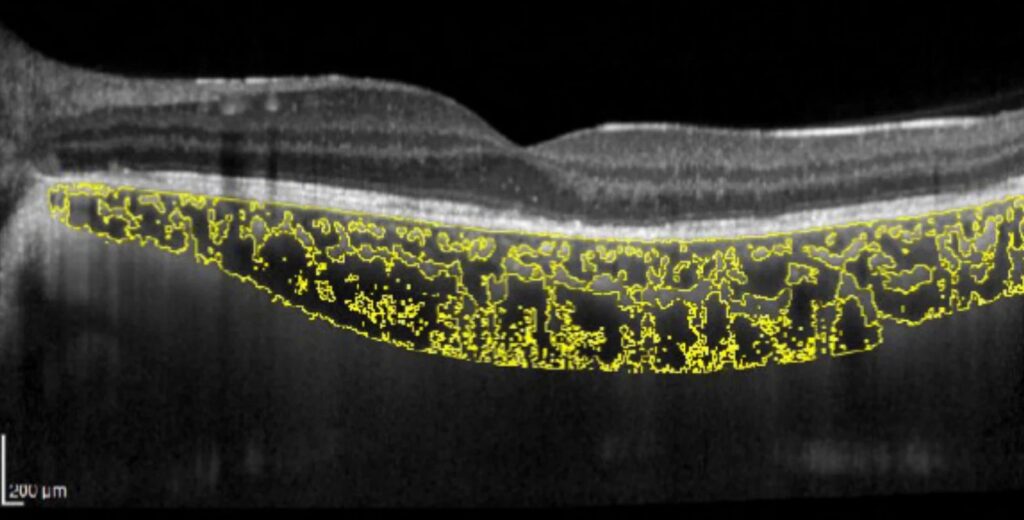
The choroidal vascularity index (CVI) is the proportion of the choroid that vessels occupy, measured on OCT images of the choroid.
Choroidal thickness changes with many factors, so researchers proposed CVI as a more stable measure of the vascular state of the choroid.
Researchers have applied it to a range of diseases, and interest in it continues to grow.
The Idea Behind the Choroidal Vascularity Index
Large and medium vessels, the choriocapillaris, and stroma make up the choroid, with melanocytes, collagen, and other cells.
Thickness is also the sum of these parts.
A thick choroid may be due to dilated vessels or to swollen stroma.
By separating the luminal area from the stromal area on a scan, CVI aims to show whether a change in thickness reflects the vessels or the surrounding tissue (see enhanced depth imaging).
Choroidal Vascularity Index: How CVI Is Calculated
- The operator obtains an OCT scan of the choroid with enhanced depth imaging or swept-source technology
- The operator outlines the choroid between the RPE-Bruch membrane complex and the chorioscleral border
- Software converts the image to a binary image, separating the dark luminal areas from the lighter stromal areas by a threshold method
- Software calculates the total choroidal area, the luminal area, and the stromal area
- The CVI is the ratio of the luminal area to the total choroidal area, expressed as a percentage
Agrawal and colleagues described the method and reported values in a population-based group of healthy eyes (see the references).
Fundus Explorer Pro
Photograph the retinal findings described here with the phone already in your pocket — 22 D optics and built-in illumination in one handheld unit.
From Choroida — the team behind this siteNormal Values
In healthy adults, the CVI is about 65 percent in many studies, with a range of several percentage points.
It varies much less between individuals than thickness.
The value changes little with the time of day, in some studies.
Reports describe it as relatively stable with age compared with thickness, though findings vary between reports.
Different devices, software, and thresholds give different numbers, so compare values across studies with caution.
Choroidal Vascularity Index: Disease Findings
- Central serous chorioretinopathy: the CVI tends to be higher, which fits with the dilated vessels and congestion of the pachychoroid spectrum (see central serous chorioretinopathy)
- Age-related macular degeneration: values are often lower in eyes with advanced disease, with some differences between types
- Diabetic retinopathy: reports show reduced or variable CVI, with changes related to severity
- Uveitis, such as Vogt-Koyanagi-Harada disease: the CVI changes with inflammation and tends to move toward normal with treatment (see Vogt-Koyanagi-Harada disease)
- High myopia: thinner choroids with a lower or normal CVI, depending on the study
- Other diseases, such as retinal vascular occlusions, glaucoma, and systemic conditions
These studies suggest that CVI may reflect the vascular state, but its meaning in an individual patient is still uncertain.
Advantages
- It is a ratio, so it is less affected by the factors that change absolute thickness, such as axial length
- You can also calculate it from existing images, without additional testing
- It may also reflect the disease process better than thickness in some conditions
- It is a possible marker of treatment response
Limitations of Choroidal Vascularity Index
- There is no standard method, and the binarization threshold is arbitrary
- The measurement depends on image quality, depth of the scan, and the segmentation of the borders
- Different devices produce different values
- Shadowing and signal falloff in deep tissue can also affect the result
- Evidence is largely from small and cross-sectional studies, and nobody has established its use in clinical decisions
Reporting of the method and the device is essential in studies.
Practical Pointers
CVI is not part of routine clinical care at present, and it is mostly a research tool.
Clinicians who see it in a report should also ask how it was calculated, and compare only values produced by the same method.
It can also add a layer of information to choroidal thickness in complex cases, and may help to separate vascular from stromal swelling.
Example of Clinical Use
In a patient with chronic central serous chorioretinopathy, the CVI may be higher than in the fellow eye, supporting a vascular cause.
It may fall after successful photodynamic therapy as the vessels become less congested.
In a patient with Vogt-Koyanagi-Harada disease, the CVI changes as the inflammation settles, and monitoring it may give a sense of whether the treatment is working.
These examples are mostly from research studies, and researchers are still establishing the value for the individual patient.
Software and Standardization
Clinicians use several software tools for binarization, and some devices have built-in programs.
Also, standardized protocols for scan acquisition and for analysis would improve comparability, and consensus papers on this topic are in development.


Document what you see
Two smartphone imaging tools built for everyday clinic use — one for the slit lamp, one for the fundus.
From Choroida — the team behind this siteReferences
- Agrawal R, Gupta P, Tan KA, et al. Choroidal vascularity index as a measure of vascular status of the choroid: measurements in healthy eyes from a population-based study. Sci Rep. 2016;6:21090.
- Iovino C, Pellegrini M, Bernabei F, et al. Choroidal vascularity index: an in-depth analysis of this novel optical coherence tomography parameter. J Clin Med. 2020;9:595.
- Ryan SJ, Schachat AP, Wilkinson CP, et al, eds. Retina. 6th ed. Elsevier; 2018.
- American Academy of Ophthalmology. Basic and Clinical Science Course, Section 12: Retina and Vitreous. AAO; current edition.

Writes and reviews the clinical articles on Educate. Every post is checked against peer-reviewed sources before it goes live.