
CASE REPORT
A 45-year-old patient presented with several notable ocular complaints in the right eye (OD), including a gradual and progressive reduction in vision over the past two months, and the presence of bothersome floaters resembling dark specks or lines in their visual field.
Moreover, sporadic mild eye pain localized to the right eye and heightened sensitivity to light (photophobia).
Upon conducting a comprehensive ocular examination, a series of significant findings were observed.
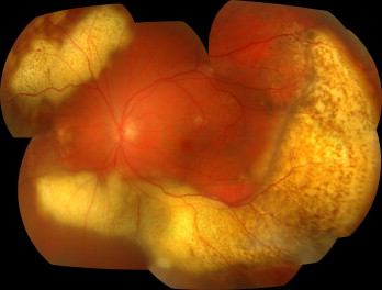
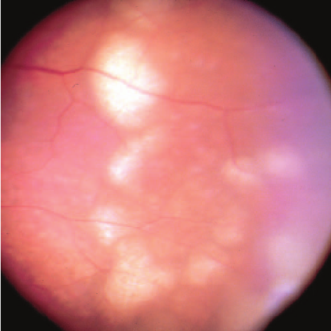
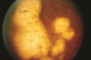
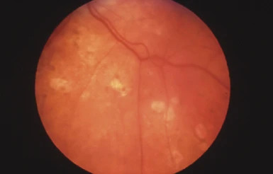
These included a marked reduction in visual acuity exclusive to the right eye (OD), the presence of mild conjunctival injection and anterior chamber cell, and multiple distinct white-yellowish subretinal infiltrates.
Also, hyperfluorescent spots were detected during fluorescein angiography, as well as the confirmation of vitreous opacities and subretinal infiltrates through B-scan ultrasonography—all of which were specific to the right eye (OD).
This complex constellation of ocular signs strongly indicated the presence of intraocular lymphoma, with the pathology predominantly localized in the right eye (OD).
Intraocular Lymphoma DISEASE entity
Primary intraocular lymphoma often poses a diagnostic dilemma with presentations like vitritis, intermediate uveitis, or subretinal plaque-like lesions.

Diagnosis is often challenging in such cases, and this is why it is often one of the diseases referred to as a masquerade syndrome.
Vitreoretinal lymphoma (as primary intraocular lymphoma is now known) is the most common intraocular lymphoproliferative disease.
The term vitreoretinal lymphoma distinguishes it from other intraocular lymphoproliferations including choroidal lymphomas (which do not have any association with central nervous system disease) and iris or ciliary body lymphomas.
The term intraocular lymphoma was first introduced more than 60 years ago. However, prior to the advent of immunohistochemistry, vitreoretinal lymphomas were known as reticulum cell sarcomas, microgliomas, perithelial sarcomas, or lymphosarcomas.
It is considered a variant of primary central nervous system (CNS) lymphomas and may occur only in the eye initially (thus a primary vitreoretinal lymphoma) or contemporaneously with CNS disease.
Rarely, a vitreoretinal lymphoma can be classified as secondary when it arises due to metastasis from a systemic lymphoma.
The majority of vitreoretinal lymphomas are of a diffuse large B-cell (DLBCL) histologic subtype, though occasionally T-cell lymphomas can occur.
Vitreoretinal lymphomas represent < 1% of all intraocular tumors, 4-6% of all intracranial tumors, and 1-2% of all extranodal non-Hodgkin’s lymphomas.
Involvement of the CNS is common, developing in 35-90% along the course of the disease. Women are more commonly affected than men and patients generally present in the 4th to 6th decade, although a case as young as 15 years has been reported.
Eighty to ninety percent of patients will have bilateral disease, although the initial presentation may be unilateral or asymmetric.
Intraocular Lymphoma MANAGEMENT
The treatment can be aimed as local therapy which can be radiotherapy to the eye or intracameral/intravitreal agents like (methotrexate and rituximab) or systemic therapy which can be external beam radiotherapy or systemic chemotherapy.
Medical therapy
EBRT(External Beam Radiotherapy)
In cases of bilaterality, EBRT is the most effective treatment. A total dose of 30-40 Gy, divided in fractions of 1.5 to 2 Gy is often used.
The side effects associated are dermatitis, punctate keratopathy, cataracts, and radiation retinopathy. The 2-year overall and disease-free survival rates were reported to be 74% and 58% respectively.
Local Chemotherapy
Methotrexate
In unilateral cases, intravitreal methotrexate has been used in the dosage of 400 µg/0.1cc twice weekly for 4 weeks, followed by 1 weekly for 4 weeks, followed by 1 monthly for 12 months.
It is used as a primary therapy as an alternative to radiotherapy or for cases of relapse. The risks associated are conjunctival injection and keratopathy.
Sometimes these can be very severe which warrants the use of alternatives. Clinical remission is achieved after a mean of 6.4 +/- 3.4 injections.
Rituximab
Intravitreal rituximab which is a chimeric anti CD20 monoclonal antibody can be used in the dosage of 1mg /0.1 ml in cases that are unresponsive or cannot tolerate methotrexate.
For isolated ocular lymphoma, local chemotherapy and or radiotherapy can be done. In cases of systemic involvement or CNS lymphoma, systemic chemotherapy with CHOP (cyclophosphamide, doxorubicin, vincristine, prednisolone) or rituximab-CHOP is done.
Other systemic agents that have been investigated include pomalidomide, stem cell transplantation, or ibrutinib, with or without local therapy.
Systemic Chemotherapy
Methotrexate
Intravenous high-dose methotrexate is commonly used in patients with intraocular lymphoma who have CNS or systemic involvement.
Rituximab
In cases with CNS involvement, rituximab may be used in conjunction with high-dose methotrexate.
Would you have interest in taking retinal images with your smartphone?
Fundus photography is superior to fundus analysis as it enables intraocular pathologies to be photo-captured and encrypted information to be shared with colleagues and patients.
Recent technologies allow smartphone-based attachments and integrated lens adaptors to transform the smartphone into a portable fundus camera and Retinal imaging by smartphone.
RETINAL IMAGING BY YOUR SMARTPHONE
REFERENCES
- Grange LK, Kouchouk A, Dalal MD, Vitale S, Nussenbla RB, Chan CC, et al. Neoplastic masquerade syndromes in patients with uveitis. Am J Ophthalmol 2014;157:526‐31.
- Biswas J, Majumdar PD .Uveitis: An Update.Goto H. Intraocular lymphoma.2016. 93-100
- Coupland SE, Damato B. Understanding intraocular lymphomas. Clin Experiment Ophthalmol. 2008;36:564-578.
- Cooper, E.L. & Riker, J.L. (1951) Malignant lymphoma of the uveal tract. American Journal of Ophthalmology, 34, 1153–1158.
- Qualman, S.J., Mendelsohn, G., Mann, R.B. & Green, W.R. (1983) Intraocular lymphomas. Natural history based on a clinicopathologic study of eight cases and a review of the literature. Cancer, 52, 878–886.
RETINAL IMAGING BY YOUR SMARTPHONE

RETINAL IMAGING BY YOUR SMARTPHONE