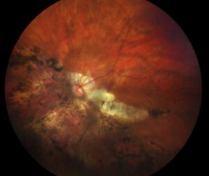
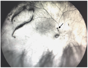
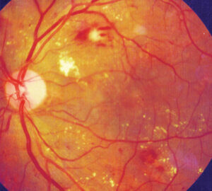
Vitreous structure is a delicate network of collagen and hyaluronan that fills the back of the eye.
It makes up about four fifths of the eye’s volume.
Therefore, its changes with age shape many common retinal disorders.
What Vitreous Structure Is
The vitreous is a transparent gel of about 4 milliliters.
It is more than 98 percent water.
Also, it is attached to the retina at several sites.
As a result, it supports the eye and links to retinal disease.
Collagen Framework of Vitreous Structure
Type II collagen forms thin fibrils with types V, IX and XI.
These fibrils form a scaffold that traps water.
Also, they are kept apart by hyaluronan.
Therefore, the gel stays clear and does not collapse.
Fundus Explorer Pro
Photograph the retinal findings described here with the phone already in your pocket — 22 D optics and built-in illumination in one handheld unit.
From Choroida — the team behind this siteHyaluronan and Other Components
Hyaluronan is a large sugar polymer that binds water and fills spaces between fibrils.
Proteoglycans such as chondroitin sulfate and opticin help to organize the network.
Also, the vitreous contains ascorbate at high concentration.
In addition, hyalocytes in the cortex produce part of these molecules.
Regions of Vitreous Structure
The cortex is the outer, denser layer near the retina.
The core is more fluid and contains the central channel known as Cloquet’s canal.
Also, the base straddles the ora serrata and is the firmest attachment.
Therefore, surgeons treat the base with care.
Sites of Firm Attachment
The vitreous attaches firmly at the base, the optic disc margin and the fovea.
It also attaches along the large retinal vessels and at lattice areas.
Also, adhesion at the macula keeps the cortex on the surface in many adults.
As a result, these sites decide where traction and tears occur.
Vitreous Structure in Youth
The young gel is homogeneous and firmly attached.
It acts as a shock absorber for the eye.
Also, it limits oxygen flow to the lens.
Therefore, removal of the gel changes the oxygen exposure of the lens (see cataract after vitrectomy).
Vitreous Structure and Liquefaction with Age
Sebag showed that the gel begins to liquefy in childhood.
By the ninth decade, more than half of the volume is liquid.
Also, collagen fibrils aggregate into thick cables.
As a result, pockets of fluid appear within the gel.
Vitreous Structure and Posterior Vitreous Detachment
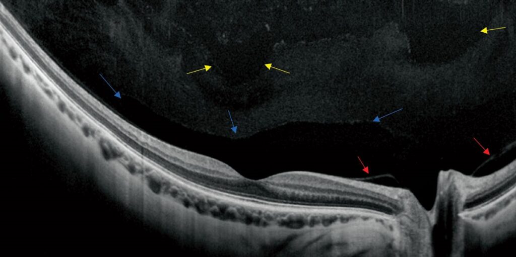
When liquid fluid enters the space behind the cortex, the gel separates from the retina.
Johnson described the stages of the process, starting at the perifoveal region.
Also, complete detachment frees the cortex from the disc.
Therefore, symptoms such as flashes and floaters follow.
Vitreoschisis
Sometimes the cortex splits into layers during detachment.
The outer layer stays on the retina and can drive membrane formation.
Also, this splitting links to macular holes and epiretinal membranes.
Therefore, it explains some of the variation in detachment outcomes.
Complications Linked to Vitreous Changes
Traction at firm sites can cause retinal tears.
Persistent attachment at the macula leads to vitreomacular traction (see vitreomacular traction).
Also, vitreous hemorrhage can follow vessel avulsion.
In addition, remaining cortex is the scaffold for proliferative disease.
Floaters and Opacities
Aggregated collagen and cells cast shadows on the retina.
Weiss rings form from the detached disc margin.
Also, asteroid hyalosis adds calcium soaps in the gel.
Therefore, floaters range from harmless to a sign of a tear.
Diabetes and Myopia
Diabetes causes glycation of collagen and earlier liquefaction.
Myopia leads to early and extensive liquefaction.
Also, both raise the risk of complicated detachment.
As a result, patients with these conditions need careful follow-up.
Role in Drug Delivery
The vitreous slows the movement of large molecules after intravitreal injection.
Vitrectomized eyes clear drugs faster.
Also, gel-like formulations aim to use this property.
Therefore, vitreous state affects drug duration.
Surgical Implications
Pharmacologic vitreolysis tried to induce detachment with enzymes.
Ocriplasmin showed partial success in selected eyes.
Also, vitrectomy removes the gel and its tractional scaffold.
In addition, the choice depends on the disease and the attachment.
Pitfalls to Avoid
- Do not assume that every floater is harmless
- Never skip dilated examination after a sudden onset of flashes
- Do not forget that the base cannot be peeled in surgery
- Never ignore the role of the vitreous in the choice of drug route
Hyalocytes
Hyalocytes sit in the vitreous cortex and clear debris.
They also respond to injury and may help scar formation.
Also, they produce hyaluronan and collagen in small amounts.
Therefore, they act as the resident immune cells of the gel.
Examining the Vitreous
Use a slit lamp with a wide beam or a contact lens to see the cortex.
Ultrasound shows detachment when the view is poor.
Also, OCT shows the attachment at the macula.
In addition, record the status in every retinal note.
For the latest studies, see the PubMed results for vitreous structure.


Document what you see
Two smartphone imaging tools built for everyday clinic use — one for the slit lamp, one for the fundus.
From Choroida — the team behind this siteReferences
- Sebag J. Age-related changes in human vitreous structure. Graefes Arch Clin Exp Ophthalmol. 1987;225:89-93.
- Bishop PN. Structural macromolecules and supramolecular organisation of the vitreous gel. Prog Retin Eye Res. 2000;19:323-344.
- Sebag J. Vitreous: the resplendent enigma. Br J Ophthalmol. 2009;93:989-991.
- Johnson MW. Posterior vitreous detachment: evolution and complications of its early stages. Am J Ophthalmol. 2010;149:371-382.
Test yourself
A few questions straight from this article.
-
Approximately what proportion of the vitreous gel is water?
The vitreous is a transparent gel of about 4 millilitres that is more than 98 percent water, filling about four fifths of the eye's volume. -
Which collagen type forms the main fibrils of the vitreous scaffold, together with types V, IX and XI?
Type II collagen forms thin fibrils with types V, IX and XI, creating a scaffold that traps water. -
Which vitreous component keeps the collagen fibrils apart so the gel stays clear and does not collapse?
Hyaluronan is a large water-binding sugar polymer that fills the spaces between fibrils and keeps them apart, maintaining a clear gel. -
Which region of the vitreous is its firmest attachment and cannot be peeled during surgery?
The vitreous base straddles the ora serrata and is the firmest attachment, which is why surgeons treat it with care and do not peel it. -
By the ninth decade of life, how much of the vitreous volume has typically become liquid?
Liquefaction begins in childhood, and by the ninth decade more than half of the volume is liquid as collagen fibrils aggregate into thick cables. -
Where does posterior vitreous detachment begin in the staged process described by Johnson?
PVD starts in the perifoveal region; complete detachment later frees the cortex from the disc, producing flashes and floaters. -
In vitreoschisis, why is the outer cortical layer that stays on the retina clinically important?
When the cortex splits, the outer layer left on the retina can drive membrane formation, linking vitreoschisis to macular holes and epiretinal membranes. -
A Weiss ring floater after posterior vitreous detachment arises from which attachment site?
Weiss rings form from the vitreous detached at the disc margin, one of several opacities that cast shadows on the retina. -
How does diabetes alter the vitreous gel?
Diabetes glycates vitreous collagen and brings liquefaction forward, raising the risk of complicated detachment, as myopia also does. -
How does prior vitrectomy affect drugs given by intravitreal injection?
The vitreous gel slows the movement of large molecules after injection, so vitrectomized eyes clear drugs faster and drug duration is shorter.

Writes and reviews the clinical articles on Educate. Every post is checked against peer-reviewed sources before it goes live.