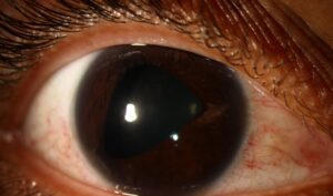
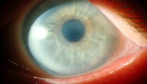
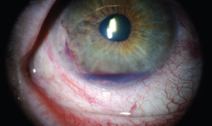
The ocular sequelae of Stevens-Johnson syndrome and toxic epidermal necrolysis often matter more to a patient’s long-term vision than the acute illness itself, since the ocular surface damage from the acute drug reaction can continue to progress silently for years afterward through chronic inflammation and scarring, long after the skin has healed and the patient may consider themselves recovered.
The Acute Phase
Stevens-Johnson syndrome and toxic epidermal necrolysis are severe, usually drug-induced, mucocutaneous reactions causing widespread epidermal and mucosal necrosis, including the ocular surface in a substantial proportion of affected patients.
Acute ocular involvement ranges from mild conjunctival injection to severe pseudomembranous conjunctivitis with corneal epithelial defects, and the severity of acute involvement is one of the strongest predictors of chronic ocular sequelae, though even patients with modest acute involvement can develop significant late complications.
Why Aggressive Acute Treatment Matters
Amniotic membrane transplantation applied to the ocular surface during the acute phase, within the first days to roughly two weeks of onset, has substantially reduced the rate of severe chronic ocular sequelae in reported series compared with supportive care alone.
This reflects a broader principle in Stevens-Johnson syndrome ophthalmic care: the chronic scarring and limbal stem cell failure that develop later are, in large part, the consequence of the acute inflammatory and cicatrizing process, so intervening early and aggressively during the acute phase has a real chance of changing the entire long-term trajectory for that patient’s eyes.
All-fit Slit-Lamp Adapter
Record and share exactly what you see at the slit lamp. One adapter fits any slit lamp or surgical microscope — and any smartphone.
From Choroida — the team behind this siteChronic Ocular Sequelae
- Dry eye disease, from destruction of the lacrimal gland ducts, goblet cells, and meibomian glands during the acute illness, often severe and lifelong
- Symblepharon, adhesions between the palpebral and bulbar conjunctiva, which can progressively restrict eye movement and eyelid function
- Trichiasis and entropion, from eyelid margin scarring, causing chronic mechanical trauma to the cornea from misdirected lashes
- Limbal stem cell deficiency, from destruction of the limbal stem cell niche during the acute inflammatory process, leading to progressive corneal conjunctivalization, vascularization, and opacification (see limbal stem cell deficiency)
- Corneal scarring and, in severe cases, keratinization of the ocular surface
- Meibomian gland dysfunction and loss, contributing further to the severe evaporative and cicatricial dry eye picture
Long-Term Surveillance
Patients who have had Stevens-Johnson syndrome or toxic epidermal necrolysis with ocular involvement need lifelong ophthalmic follow-up, since progressive scarring and surface failure can continue to develop years after the acute illness, even when the eyes appeared relatively stable in the interim.
- Regular assessment of eyelid margin position and lash direction, since trichiasis can develop or progress gradually
- Monitoring for symblepharon progression and fornix shortening
- Assessment of corneal clarity and limbal stem cell function over time
- Aggressive, ongoing management of the severe chronic dry eye that most patients develop
Management of Chronic Disease
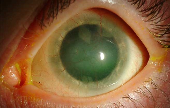
Ocular Surface Support
Intensive, preservative-free lubrication, punctal occlusion, and scleral contact lenses, which vault over the irregular and dry corneal surface and bathe it in a fluid reservoir, are central to managing chronic dry eye and can substantially improve both comfort and vision in appropriately fitted patients.
Eyelid Surgery
Trichiasis and entropion require surgical correction, sometimes repeatedly over time as scarring continues, to prevent ongoing mechanical corneal trauma.
Limbal Stem Cell and Corneal Surgery
Standard limbal stem cell transplantation is often less successful in Stevens-Johnson syndrome than in other causes of limbal stem cell deficiency, because the ongoing inflammatory and cicatrizing surface environment tends to cause transplanted stem cells to fail; for this reason, keratoprosthesis, particularly the Boston type II or osteo-odonto-keratoprosthesis, is more often considered for severe bilateral disease with an inhospitable ocular surface (see Boston keratoprosthesis).
Prognosis
Ocular outcomes vary enormously, from patients with mild, well-controlled chronic dry eye and normal vision to those with severe, progressive surface failure and significant visual impairment despite aggressive treatment.
Early, aggressive acute-phase treatment, particularly with amniotic membrane transplantation when indicated, along with vigilant, lifelong follow-up for chronic sequelae, offers the best chance of preserving vision over the patient’s lifetime.


Document what you see
Two smartphone imaging tools built for everyday clinic use — one for the slit lamp, one for the fundus.
From Choroida — the team behind this siteReferences
- Gregory DG. Treatment of acute Stevens-Johnson syndrome and toxic epidermal necrolysis using amniotic membrane: a review of 10 consecutive cases. Ophthalmology. 2011;118:908-914.
- Sotozono C, Ueta M, Koizumi N, et al. Diagnosis and treatment of Stevens-Johnson syndrome and toxic epidermal necrolysis with ocular complications. Ophthalmology. 2009;116:685-690.
- Jain R, Sharma N, Basu S, et al. Stevens-Johnson syndrome: the role of an ophthalmologist. Surv Ophthalmol. 2016;61:369-399.
- Iyer G, Pillai VS, Srinivasan B, et al. Mucous membrane grafting for lid margin keratinization in Stevens-Johnson syndrome. Cornea. 2010;29:145-151.
Test yourself
A few questions straight from this article.
-
Stevens-Johnson syndrome and toxic epidermal necrolysis are usually induced by which exposure?
Both are severe, usually drug-induced mucocutaneous reactions that cause widespread epidermal and mucosal necrosis, including the ocular surface. -
Why do patients recovered from Stevens-Johnson syndrome with eye involvement need lifelong ophthalmic review?
Cicatrisation and ocular surface failure can keep progressing years after the acute illness, even when the eyes looked stable in the interim. -
Amniotic membrane transplantation for acute Stevens-Johnson syndrome is applied within which window from onset?
Acute-phase amniotic membrane applied within the first days to about two weeks has reduced severe chronic sequelae compared with supportive care alone. -
Why does aggressive acute-phase ocular treatment in Stevens-Johnson syndrome alter the long-term visual trajectory?
The chronic scarring and limbal stem cell failure seen later are largely a consequence of the acute inflammatory and cicatrising phase, so early intervention can change the whole course. -
Severe chronic dry eye after Stevens-Johnson syndrome follows acute destruction of which structures?
The acute illness destroys lacrimal gland ducts, goblet cells and meibomian glands, producing dry eye that is often severe and lifelong. -
Symblepharon after Stevens-Johnson syndrome describes adhesions between which two surfaces?
Symblepharon is adhesion between the palpebral and bulbar conjunctiva, and it can progressively restrict eye movement and eyelid function. -
Limbal stem cell deficiency after Stevens-Johnson syndrome produces which corneal change?
Destruction of the limbal stem cell niche leads to progressive corneal conjunctivalisation, vascularisation and opacification. -
How do scleral contact lenses help the chronic ocular surface in Stevens-Johnson syndrome?
Scleral lenses vault over the irregular, dry cornea and bathe it in a fluid reservoir, improving both comfort and vision in well-fitted patients. -
Why is standard limbal stem cell transplantation often unsuccessful in Stevens-Johnson syndrome?
The persistently inflammatory and cicatrising surface environment tends to make transplanted limbal stem cells fail, unlike other causes of limbal deficiency. -
Which option is more often considered for severe bilateral Stevens-Johnson syndrome with an inhospitable ocular surface?
Because limbal grafts tend to fail on such a surface, keratoprosthesis, particularly Boston type II or osteo-odonto-keratoprosthesis, is favoured in severe bilateral disease.

Writes and reviews the clinical articles on Educate. Every post is checked against peer-reviewed sources before it goes live.