
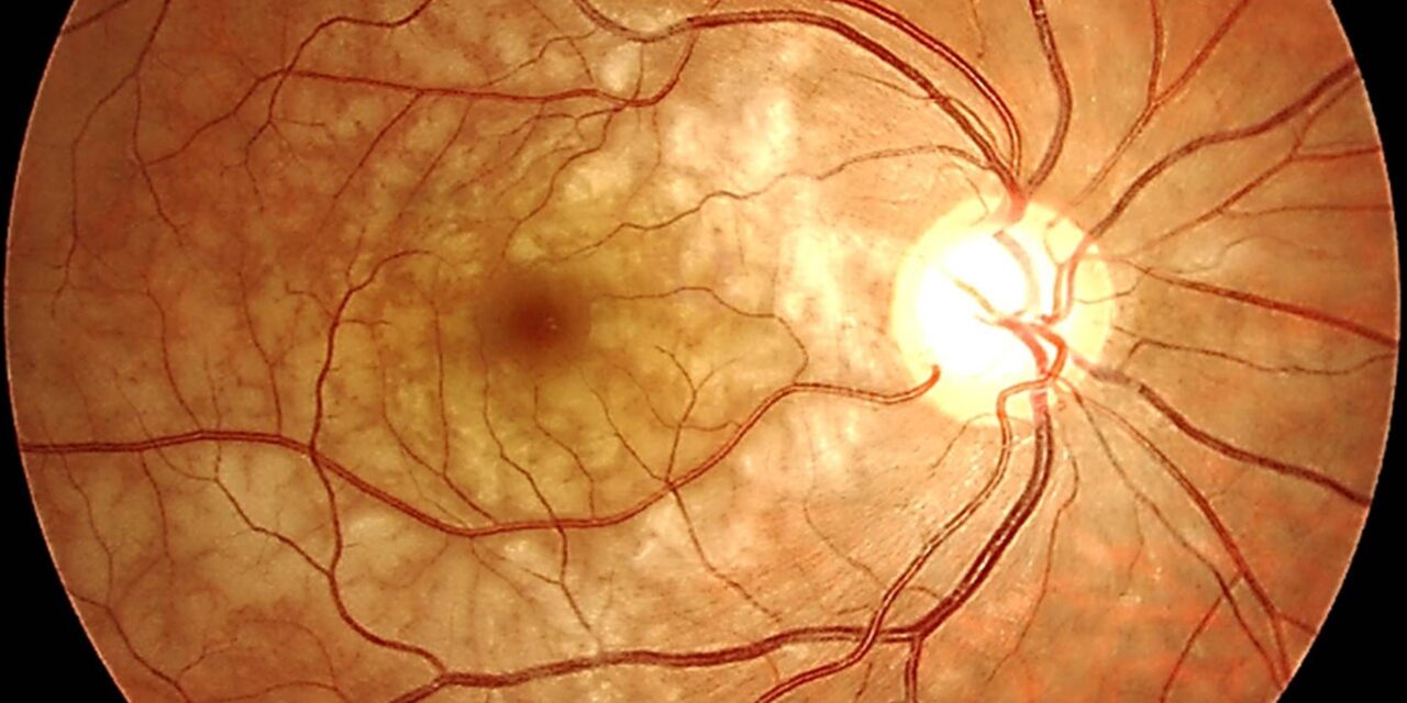
Paracentral acute middle maculopathy (PAMM) is a retinal finding defined almost entirely by its OCT appearance — a hyperreflective band at the level of the inner nuclear layer, representing infarction of the intermediate retinal capillary plexus — and it is a useful example of how OCT has changed clinical practice.
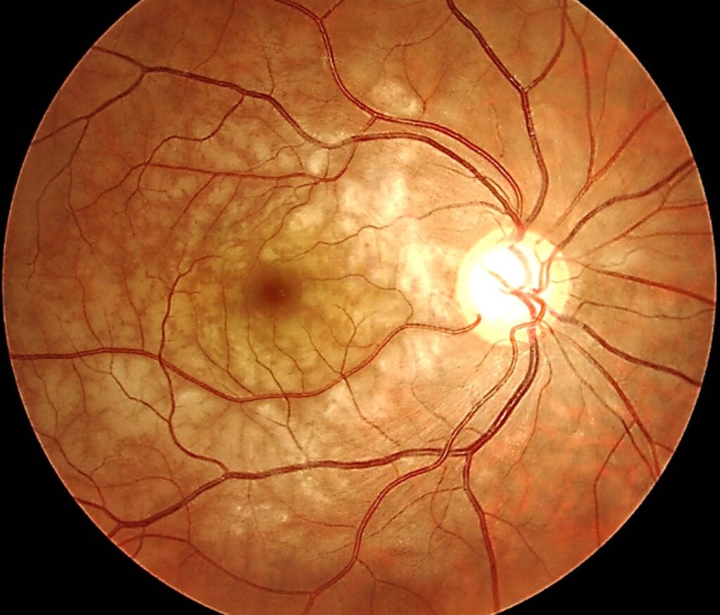
This is because PAMM is frequently subtle or entirely invisible on standard fundus examination, meaning the diagnosis is often made only because OCT was obtained and specifically, carefully reviewed.
Pathophysiology
The retina’s middle capillary plexus, positioned between the more familiar superficial and deep capillary networks, supplies the inner nuclear layer.
Ischemia specifically affecting this watershed-type vascular territory produces the characteristic band of hyperreflectivity and, eventually, thinning at that level on OCT.
This is mechanistically related to, but distinct from, the more familiar acute macular neuroretinopathy (AMN), which affects the outer retina at the level of the outer nuclear layer and outer plexiform layer rather than the inner nuclear layer.
The two conditions are now understood to represent ischemia at different depths of the retinal capillary network, sometimes occurring together in the same patient.
Clinical Presentation
Patients report the acute or subacute onset of a paracentral scotoma — a blind or dim spot near, but typically not involving, the center of fixation — sometimes accompanied by mild blurring.
However, visual acuity itself may be relatively preserved depending on how close the lesion sits to the fovea.
Symptoms can be subtle enough that some patients do not seek care immediately.
The finding is sometimes discovered only when imaging is obtained for another reason or during workup of a broader vascular event.
Because the visual symptoms can be so mild and easy to attribute to something else, a patient’s own description of a subtle, persistent dim area near — but not exactly at — the center of vision should be taken seriously and specifically prompt macular OCT, even when a cursory fundus exam looks entirely unremarkable.
Fundus Explorer Pro
Photograph the retinal findings described here with the phone already in your pocket — 22 D optics and built-in illumination in one handheld unit.
From Choroida — the team behind this siteAssociated Conditions
- Retinal vascular occlusive disease — branch or central retinal artery or vein occlusion, with PAMM sometimes representing a milder, more localized ischemic event either preceding, accompanying, or following a larger occlusive event
- Systemic vascular risk factors — hypertension, diabetes, and other conditions predisposing to retinal microvascular disease, similar to the risk factor profile for retinal vein and artery occlusions generally
- Migraine — PAMM has been reported in association with migraine, particularly migraine with aura, in an otherwise low-risk population
- Sickle cell disease and other hemoglobinopathies
- Systemic conditions causing widespread vascular stress — severe anemia, hypotensive episodes, and other causes of compromised retinal perfusion
- Idiopathic cases, without an identifiable systemic trigger, particularly in younger patients
Diagnostic Evaluation
OCT is essential and, in practice, is what actually makes the diagnosis, showing the characteristic hyperreflective band at the inner nuclear layer level acutely.
This evolves into inner nuclear layer thinning and atrophy over subsequent weeks to months as the affected tissue undergoes permanent structural loss.
Because PAMM is so often a marker of an underlying retinal vascular event, a search for the broader occlusive process — careful fundus exam, OCT angiography where available to assess the retinal capillary plexuses directly, and fluorescein angiography in select cases — is an important part of the workup, rather than treating the PAMM finding as an isolated, self-contained diagnosis.
Given the association with systemic vascular risk factors, a reasonable baseline evaluation includes blood pressure measurement, screening for diabetes, and consideration of a broader vascular risk factor assessment, particularly in patients without an obvious alternative explanation such as migraine, treating the PAMM finding as an entry point into a broader systemic vascular risk assessment rather than a purely ophthalmic curiosity.
Differential Diagnosis
- Acute macular neuroretinopathy — outer retinal (outer nuclear/outer plexiform layer) involvement rather than inner nuclear layer, with its own dedicated coverage on this site, sometimes coexisting with PAMM in the same eye
- Cotton wool spots — a more familiar, related finding of focal nerve fiber layer infarction, visible on fundus exam (unlike PAMM, which often is not) and representing ischemia at a different, more superficial retinal level
- Branch retinal artery occlusion — a larger, more extensive area of retinal whitening and ischemia, generally visible on fundus exam, representing occlusion of a larger vessel rather than the capillary-level ischemia of PAMM
Management and Prognosis
There is no specific, proven treatment that reverses established PAMM.
Management is directed at identifying and addressing any underlying systemic vascular risk factor or occlusive process, in the same way any other manifestation of retinal vascular disease would be approached, with the systemic workup generally taking priority over any direct attempt to treat the retinal finding itself.
The paracentral scotoma associated with PAMM is often permanent, corresponding to the area of permanent inner nuclear layer thinning that develops as the acute finding resolves.
However, the functional impact varies considerably depending on lesion size and proximity to the fovea, and some patients experience meaningful subjective improvement over time despite persistent structural change on OCT, likely reflecting some degree of neural and perceptual adaptation to the fixed structural deficit rather than true anatomic recovery.


Document what you see
Two smartphone imaging tools built for everyday clinic use — one for the slit lamp, one for the fundus.
From Choroida — the team behind this siteReferences
- Sarraf D, Rahimy E, Fawzi AA, et al. Paracentral acute middle maculopathy: a new variant of acute macular neuroretinopathy associated with retinal capillary ischemia. JAMA Ophthalmology.
- Rahimy E, Kuehlewein L, Sadda SR, Sarraf D. Paracentral acute middle maculopathy: what we knew then and what we know now. Retina.
- American Academy of Ophthalmology. Basic and Clinical Science Course, Section 12: Retina and Vitreous.
