Disease
Idiopathic retinal vasculitis, aneurysms, and neuroretinitis (IRVAN) syndrome is a rare clinical entity of unknown etiology.
Some authors have speculated an association between positive P-ANCA s (Perinuclear AntiNeutrophil Cytoplasmic Antibodies) systemic vasculitis and antiphospholipid syndrome.
Stages
- Stage 1: Macroaneurysms, exudation, neuroretinitis, retinal vasculitis.
- Stage 2: Capillary nonperfusion (angiographic evidence)
- Stage 3: Posterior segment neovascularization of disc or elsewhere and/or vitreous hemorrhage.
- Stage 4 : Anterior segment neovascularization (rubeosis iridis)
- Stage 5 : Neovascular glaucoma.
Fundus Explorer Pro
Photograph the retinal findings described here with the phone already in your pocket — 22 D optics and built-in illumination in one handheld unit.
From Choroida — the team behind this siteDiagnosis
The diagnosis of Idiopathic retinal vasculitis, aneurysms, and neuroretinitis (IRVAN) is based on a constellation of clinical features.
Three major criteria (retinal vasculitis, aneurysmal dilations at arterial bifurcations, and neuroretinitis) and 3 minor criteria (peripheral capillary nonperfusion, retinal neovascularization, and macular exudation) are used to diagnose IRVAN.
Patients with IRVAN usually present with decreased visual acuity in one or both eyes. They may have a variable number of cells in the anterior chamber and the vitreous.
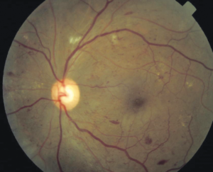
The retinal arterioles have multiple aneurysmal dilatations extending through the first several orders of arterial branching until mid-periphery.
The aneurysms are commonly found on the optic nerve, a site where typical senile retinal macroaneurysms are not. The aneurysms are typically found at branching points of the arteries, and may be fusiform, Y-shaped, or spheroid in shape.
In segments between the aneurysms the retinal arteries frequently show marked caliber variations. Arterial aneurysms clinically resemble multiple and consecutive “knots of a cord”.
Disc neovascularization may be observed in severe and advanced cases of IRVAN, with peripheral capillary dropout. Vitreous hemorrhage can result from retinal neovascularization.
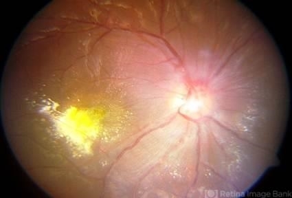
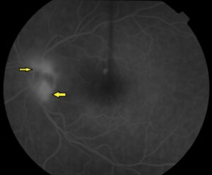
Non-complicated cases may have a slight swelling of the optic nerve head, highlighted in the late phases of fluorescein angiography.
Some authors think there are no true neuroretinitis, leakage occurs from the aneurysms on the optic nerve head, and periarterial lipid exudation results from retinal artery aneurysms.
Retinal vascular sheathing may occur, but it is not a prominent feature of the disorder in most patients. In the retinal periphery, there is a variable amount of retinal nonperfusion, with obliteration of the arteries, veins, and retinal capillaries.
In the perfused area bordering the peripheral areas of nonperfusion, the retinal veins may show abnormal caliber alterations.
If the ischemic load of the nonperfused retina reaches a critical level, retinal and iris neovascularization may develop. Interestingly, neo-vessels may sometimes reproduce arterial macroaneurysms seen in the retinal vasculature ( Richard F Spaide MD, personal communication ).
In severely affected individuals, vitreous hemorrhage and neovascular glaucoma have developed.
Although lipid deposition is not common in most patients with uveitis, patients with IRVAN frequently have marked intraretinal lipid deposition.
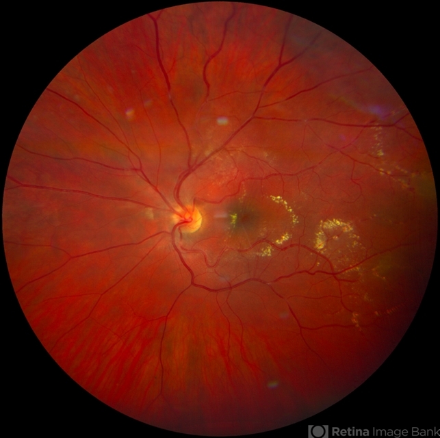
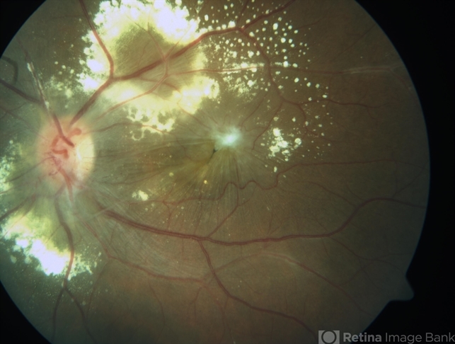
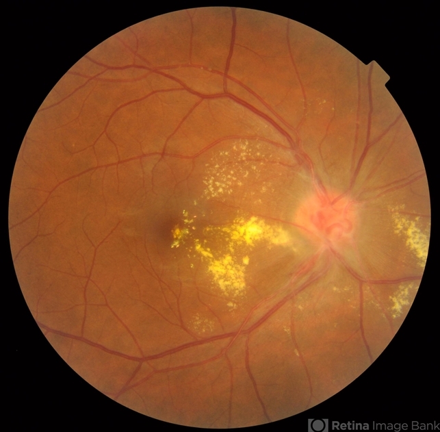
This lipid is usually found in the peripapillary region and may extend into the macula leaving a macular star or massive lipid accumulation.
The lipid is generally found near the greatest concentration of aneurysms ( peripapillary location and along the arcades ).
The retina in these areas is commonly edematous and may be detached by exudative fluid. As time goes by, a fibrous grayish macular scar may develop as a legacy of long-standing exudation in the macula.
Signs
- Multiple post-equatorial arterial aneurysms in the retina and optic disc head.
- Peripapillary lipid exudation associated with macroaneurysms and retinal vasculitis.
- Retinal hemorrhages in the posterior pole.
- Peripheral capillary nonperfusion
- Retinal vascular sheathing
- Swelling of the optic nerve
- Retinal neovascularization
- Optic disc neovascularization
Symptoms
Asymptomatic
Patients may not complain about loss of vision in spite of having a severe disease if it does not affect the macula at the initial presentation.
Nonspecific symptoms
For instance, multiple visits for a change of glasses due to blurred vision.
Clinical diagnosis
Patients with Idiopathic retinal vasculitis, aneurysms, and neuroretinitis (IRVAN) are, sometimes, over-investigated for systemic causes of retinal disease.
However, IRVAN seems to produce only ophthalmic manifestations, with general workup being mostly unremarkable.
Retinal imaging is a key element for a rapid diagnosis of IRVAN, and a multimodal imaging approach is recommended when this syndrome is suspected, to collect all possible diagnostic criteria.
Dye-mediated angiographies ( fluorescein and indocyanine green) remain the standard imaging modalities for confirming the clinical diagnosis of IRVAN.
Diagnostic procedures
Fluorescein angiography:
Fluorescein angiography highlights arteriolar abnormalities. The aneurysms are more evident and the alterations in arteriolar caliber are more obvious.
Leakage of fluorescein from the aneurysms is apparent. Patients with IRVAN usually have optic nerve head staining. Of course, disc and/or retinal neovascularization shows marked fluorescein leakage.
The areas of peripheral non-perfusion are easier to appreciate with fluorescein angiography, especially wide-field fluorescein angiography.
Indocyanine green angiography:
The aneurysmal dilatations showed uniform staining. No abnormalities were seen in the choroidal vessels; in particular, no aneurysms of the choroidal vessels have been observed.
For example, the retinal abnormalities and macroaneurysms in IRVAN are more clearly imaged with ICG angiography, especially at the very late stages, but the leakage, ischemia, and retinal neovascularization are still best delineated with FA.
Optical coherence tomography:
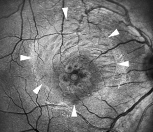
It is useful to assess macular edema, vitreoretinal traction over both the macula and optic nerve head, neovascular tufts in the posterior pole and epiretinal membrane, as an example:
Optical coherence tomography angiography ( OCT-A ):
OCT-A is a dye-free, noncontact modality to image the eye. It is a noninvasive imaging modality that provides three-dimensional images of the retina and choroidal microvasculature in vivo.
OCT-A has its clinical shortcomings and is less useful for identification of retinal vasculitis
- It doesn’t show leakage
- Patients with poor fixation are difficult to image effectively.
- Opaque media such as vitreous hemorrhage blocks the OCT signal and decreases the quality of the angiography.
- Although widefield OCT-A will allow for imaging of the vascular layers, likely out to the mid-periphery and vortex veins, ultra-widefield FA will still provide information about retinal capillary perfusion out to the ora serrata.
Differential diagnosis
At presentation, the physician must distinguish clinical elements of this interesting disease: arterial macroaneurysms, lipid exudation, retinal periphery capillary dropouts, and neovascularization.
The syndrome’s striking ophthalmoscopic picture is not shared by any other disease. However, four principal conditions may be diagnosed initially.
The first is idiopathic retinal vasculitis if the aneurysmal dilations are not noticed. Careful ophthalmoscopic and fluorescein angiographic examination should reveal any aneurysmal dilations, if present.
The second is the senile retinal arterial macroaneurysm. This acquired macroaneurysm is usually found in older patients with a history of high blood pressure and/or cardiovascular diseases. It usually is single, mostly unilateral, located temporal to the optic nerve along the midsegment of an artery, and associated with hemorrhage.
Although there may be capillary telangiectasis surrounding the macroaneurysm, there is no peripheral nonperfusion or signs of inflammation.
Patients with IRVAN are otherwise healthy young people and arterial macroaneurysms are bilateral and multiple. The third element in the differential diagnosis is Coats´ disease.
Patients with Coats’ disease have a unilateral presentation with no signs of intraocular inflammation. The aneurysms of Coats’ disease usually involve more than one element of the vascular circuit and may involve the arteries, capillaries, and veins.
The aneurysms in Coats’ disease do not involve arterial bifurcations and they are observed in the mid and far periphery usually surrounded by abundant circinate lipid exudation.
Finally, peripheral telangiectasias are irregular in shape compared to aneurysms. The fourth condition is Eales´ disease.
Peripheral retina non-perfusion is almost always present with retinal neovascularization and sometimes arising from the optic disc.
Nevertheless, it is not possible to observe typical fusiform or “tied-knot ” arterial aneurysms from IRVAN syndrome.
Management
Currently, retinal laser photocoagulation and the use of intravitreal injections of anti-vascular endothelial growth factor antibodies (anti-VEGF ) are considered within the first line of treatment.
Interestingly, arterial macroaneurysms may regress or vanish after treatment or during follow-up.
General treatment
- Observation: Basha et al have published a pediatric case with IRVAN, with one eye treated and the other eye untreated. A long-term follow-up demonstrated that observation is a consideration in IRVAN syndrome if the vision remains good with hard exudation.
Medical therapy
Corticosteroids:
Patients affected with IRVAN have been treated with corticosteroids in an attempt to reduce any inflammatory worsening of their ophthalmoscopic appearance.
Unfortunately, corticosteroids have not been particularly useful. Although corticosteroids undoubtedly decrease inflammation, much of the morbidity of IRVAN results from chronic secondary changes such as persistent leakage from the aneurysms and vitreous hemorrhage from the neovascularization.
- Systemic use: Prednisone 1mg/kg daily or intravenous
- Intravitreal injections: Triamcinolone Acetate and Dexamethasone implant to treat chronic macular edema.
Anti-VEGF intravitreal injections:
Pegaptanib ( MACUGEN ), Bevacizumab ( AVASTIN ), Ranibizumab ( LUCENTIS ) , and Aflibercept ( EYLEA ) have been used for treating IRVAN syndrome in patients with macular edema, retinal ischemia and neovascularization of the eye.
Immunosuppressive drugs:
Cheema et al and Samalia et al showed improvement in two patients treated with infliximab, an anti-TNF. TNFα might be a causative factor in mediating inflammation and causing tissue destruction in IRVAN patients.
Saatchi et al used oral azathioprine as an adjuvant in a patient with IRVAN. Ameratunga et al used monthly cycles of pulsed intravenous methylprednisolone 1 gm on 3 consecutive days for 7 months along with mycophenolate mofetil and oral prednisone 10 mg daily.
Medical follow up
Follow-up depends on the clinical condition. IRVAN patients should be followed at least every 3 months until stabilization.
Surgery
Thermal Focal Laser and Peripheral Retinal Photocoagulation:
Focal laser treatment to the macroaneurysms may decrease the amount of lipid exudation but carries the risk of causing focal occlusion of the retinal arteriole, thus its use is controversial.
Laser spots may be applied surrounding macroaneurysm areas but never directly. Laser-induced arteriolar occlusion has caused some patients to lose substantial visual acuity.
Panretinal photocoagulation to the peripheral areas of nonperfusion is performed in patients even before retinal/optic disc neovascularization appears.
Several laser sessions may be required for each eye. The aim of this treatment is to cause a regression of the new vessels. Some patients with substantial amounts of vitreous hemorrhage have required vitrectomy and laser endo photocoagulation to treat bleeding areas of retinal neovascularization.
Although retinal and vitreous hemorrhages occur from typical senile retinal arterial macroaneurysms, bleeding from the aneurysmal dilations of IRVAN is not common.
Peripheral cryotherapy:
Some authors have used this procedure, especially when opaque media is present and peripheral laser treatment cannot be applied.
Pars Plana Vitrectomy
Indications:
- Vitreous Hemorrhage that persists for more than 1-2 months.
- Dense vitreous hemorrhage (intravitreal blood/clots do not allow to see optic nerve head and/or retina details.
- Severe loss of vision secondary to vitreous hemorrhage which does not allow for treating a patient.
- Tractional retinal detachment, due to proliferative retinopathy, especially if the center of the retina (macula ) is off. Severe cases may need lensectomy, core vitrectomy, separation of posterior hyaloid, peeling of epiretinal membranes and vitreous base for eliminating vitreoretinal traction, and silicone oil for a long-term vitreous tamponade.


Document what you see
Two smartphone imaging tools built for everyday clinic use — one for the slit lamp, one for the fundus.
From Choroida — the team behind this siteREFERENCES
- Chang TS, Aylward GW, Davis JL, et al. Idiopathic retinal vasculitis, aneurysms, and neuro-retinitis. Ophthalmology. 1995;102:1089–1097.
- Samuel MA, Equi RA, Chang TS, et al. Idiopathic retinitis, vasculitis, aneurysms, and neuroretinitis (IRVAN): new observations and a proposed staging system. Ophthalmology. 2007;114:1526–1529.
- Centers for Disease Control (CDC). International Classification of Diseases, Tenth Revision, Clinical Modification(ICD-10-CM): FY 2017 version; 2017. Available at: https://www.cdc.gov/nchs/icd/icd10cm.htm. Accessed November 23,2017.
- Flynn J, Clarkson J, Curtin V, Flynn Jr. H, Hurtes R, Smith J. J. Donald M Gass, MD : Festschrift Editorial. Am J Ophthalmol 2004;137,3:480-2.
- Gass JD. Stereoscopic Atlas of Macular Diseases: Diagnosis and Treatment. 3rd ed. St Louis: Mosby; 1987.
- Basha M, Brown GC, Palombaro G, Shields CL, Shields JA. Management of IRVAN syndrome with observation. Ophthalmic Surg Lasers Imaging Retina. 2014 May 1;45.
- Krishnan R, Shah P, Thomas D. Subacute idiopathic vasculitis, aneurysms and neuroretinitis (IRVAN) in a child and review of paediatric cases of IRVAN revealing preserved capillary perfusion as a more common feature. Eye (Lond). 2015;29:145–151.
- DiLoreto DA Jr, Sadda SR. Idiopathic retinal vasculitis, aneurysms, and neuroretinitis (IRVAN) with preserved perfusion. Retina. 2003;23:554–557.
- Ishikawa F, Ohguro H, Sato S, Sato M, Yamazaki H, Nakazawa M.A case of idiopathic retinal vasculitis, aneurysm, and neuroretinitis effectively treated by steroid pulse therapy. Jpn J Ophthalmol. 2006 Mar-Apr;50(2):181-5.
- Empeslidis T, Banerjee S, Vardarinos A, Konstas AG. Dexamethasone intravitreal implant for idiopathic retinal vasculitis, aneurysms, and neuroretinitis. Eur J Ophthalmol. 2013;23:757–760.
- Mitry D, Schmoll C, Hegde V, Borooah S, Singh J, Bennett H.Use of pegaptanib in the treatment of vitreous haemorrhage in idiopathic retinal vasculitis. Eye (Lond). 2008 Nov;22(11):1449-50.
Test yourself
A few questions straight from this article.
-
Which triad makes up the three major diagnostic criteria of IRVAN syndrome?
IRVAN is diagnosed on a constellation of features whose three major criteria are retinal vasculitis, aneurysmal dilations at arterial bifurcations and neuroretinitis. -
Which three findings constitute the minor diagnostic criteria of IRVAN syndrome?
Alongside the three major criteria, IRVAN has three minor criteria: peripheral capillary nonperfusion, retinal neovascularisation and macular exudation. -
Which aneurysm location helps separate IRVAN from typical senile retinal macroaneurysms?
In IRVAN aneurysms are commonly found on the optic nerve, a site where typical senile retinal macroaneurysms are not. -
What shapes do the arterial aneurysms of IRVAN typically take?
The aneurysms sit at arterial branching points and may be fusiform, Y-shaped or spheroid, resembling consecutive knots of a cord. -
Stage 2 of the IRVAN staging system is defined by which finding?
Stage 2 is angiographic evidence of capillary nonperfusion, following the stage 1 findings of macroaneurysms, exudation, neuroretinitis and vasculitis. -
Which development marks stage 4 of IRVAN syndrome?
Stage 4 is anterior segment neovascularisation, that is rubeosis iridis; neovascular glaucoma defines stage 5. -
Where is the marked intraretinal lipid of IRVAN usually deposited?
Lipid collects where aneurysms are most concentrated, peripapillary and along the arcades, and may extend into the macula as a macular star. -
What may eventually develop in the macula after long-standing exudation in IRVAN?
The retina in exudative areas is oedematous and may detach, and over time a fibrous greyish macular scar is left as the legacy of chronic macular exudation. -
What does systemic investigation typically reveal in a patient with IRVAN syndrome?
Patients are sometimes over-investigated for systemic causes, yet IRVAN appears to produce only ophthalmic manifestations with a largely normal general workup. -
Which imaging modalities remain the standard for confirming a clinical diagnosis of IRVAN?
Multimodal imaging is recommended, but dye-mediated angiography with fluorescein and indocyanine green remains the standard for confirming the diagnosis.

Writes and reviews the clinical articles on Educate. Every post is checked against peer-reviewed sources before it goes live.