Superior orbital fissure syndrome is a combined palsy of cranial nerves III, IV, V1 and VI with a normal optic nerve.
Older texts call it Rochon-Duvigneaud syndrome. Because vision is spared, the pattern places the lesion at the fissure itself, and therefore a careful bedside exam predicts much of the imaging.
Anatomy of the Superior Orbital Fissure
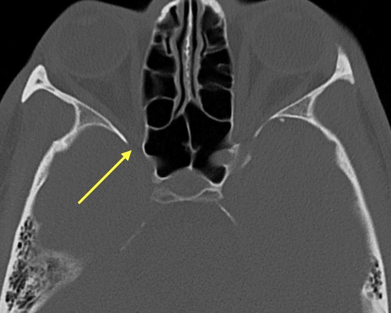
The fissure is a cleft between the greater and lesser wings of the sphenoid bone, and it links the middle cranial fossa to the orbit. Cranial nerves III, IV and VI pass through it, along with the frontal, lacrimal and nasociliary branches of V1, while the superior ophthalmic vein and sympathetic fibers also use this opening. However, the optic nerve travels separately through the optic canal.
Why Vision Is Spared
The optic strut separates the optic canal from the fissure. As a result, a lesion confined to the fissure leaves acuity, color vision and fields intact, and a new relative afferent pupillary defect therefore signals spread toward the apex.
All-fit Slit-Lamp Adapter
Record and share exactly what you see at the slit lamp. One adapter fits any slit lamp or surgical microscope — and any smartphone.
From Choroida — the team behind this siteThe Annulus of Zinn in Superior Orbital Fissure Lesions
This tendinous ring divides the fissure into two compartments, with both divisions of III, the nasociliary nerve and VI running inside it. Above it lie IV, the frontal and lacrimal nerves and the superior ophthalmic vein, and therefore a small lesion may produce an incomplete pattern.
Exam Signs of Superior Orbital Fissure Syndrome
Patients usually report binocular diplopia, a drooping lid and forehead numbness, and complete ptosis can hide the diplopia until you lift the lid.
- Ptosis with limited elevation, depression and adduction
- Absent intorsion on attempted downgaze from trochlear palsy
- Abduction deficit from abducens palsy
- A dilated, poorly reactive pupil, because parasympathetic fibers travel with III
- Reduced forehead and corneal sensation
- Mild proptosis from venous congestion
- Normal acuity and no afferent defect
Corneal Sensation and the Slit Lamp
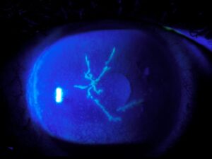
The nasociliary nerve carries corneal sensation, so test it with a cotton wisp before instilling anesthetic drops. At the slit lamp, look for punctate staining, a dull epithelium or a persistent defect, because these signs suggest early neurotrophic keratopathy, which needs lubrication and close review.
Superior Orbital Fissure Versus Orbital Apex Syndrome
Orbital apex syndrome adds optic neuropathy to the same motor and sensory deficits, so expect reduced acuity, dyschromatopsia, a field defect and an afferent pupillary defect. The disc often looks normal at first, while pallor follows weeks later, and because the two overlap, many authors treat them as one spectrum.
Superior Orbital Fissure Versus Cavernous Sinus Syndrome
The cavernous sinus can produce the same ophthalmoplegia (see cavernous sinus syndrome). However, cheek numbness from V2 points behind the fissure, because the maxillary nerve exits through foramen rotundum. A Horner syndrome with abducens palsy also favors the sinus, and bilateral signs, chemosis and fever raise concern for cavernous sinus thrombosis.
Common Causes
Trauma and tumors account for many cases, and meningioma, lymphoma, metastasis and nasopharyngeal carcinoma can all reach the fissure. Idiopathic inflammation, including Tolosa-Hunt syndrome, causes painful ophthalmoplegia, infection also matters, for example invasive fungal sinusitis or herpes zoster ophthalmicus, and finally carotid-cavernous fistula and aneurysm can mimic the syndrome.
Trauma and the Superior Orbital Fissure
Fractures of the lateral orbital wall, zygomatic complex or sphenoid can narrow the fissure, and bone fragments, hematoma and edema then compress the nerves. Thin-slice CT shows the fracture pattern and any displaced fragment, and you should always assess the optic nerve, because traumatic optic neuropathy changes urgency.
Imaging the Superior Orbital Fissure
Contrast MRI of the orbits and cavernous sinus, with fat suppression, is the preferred study, while CT adds bone detail and shows sinus disease. Next, request CT or MR angiography when you suspect a fistula or aneurysm, and in diabetic or immunosuppressed patients, arrange urgent nasal endoscopy to exclude mucormycosis.
Treatment of Superior Orbital Fissure Syndrome
Treatment targets the cause, and corticosteroids often relieve Tolosa-Hunt pain within days. However, steroids can mask lymphoma and worsen fungal infection, so exclude both first. Invasive fungal disease needs antifungals and surgical debridement, while for trauma, high-dose steroids and decompression appear in case series, although controlled evidence is lacking.
Counseling the Patient
Explain that the eye is healthy, while the nerves that move it are affected. In traumatic series, many patients regain function over months, although recovery is often incomplete, and occlusion or a temporary prism controls diplopia meanwhile. Ask the patient to return at once if vision blurs or color fades.
Follow-Up in Superior Orbital Fissure Syndrome
At each visit, measure acuity, color vision and pupils to detect apex spread, and next record ductions and a cover test to document recovery. Strabismus or ptosis surgery waits until measurements stay stable for several months.
Pitfalls to Avoid
- Do not label a pupil-involving third nerve palsy as microvascular
- Never start steroids before excluding infection and lymphoma
- Do not miss V2 numbness, because it moves the lesion to the cavernous sinus
- Avoid anesthetic drops before testing corneal sensation


Document what you see
Two smartphone imaging tools built for everyday clinic use — one for the slit lamp, one for the fundus.
From Choroida — the team behind this siteReferences
- Lenzi GL, Fieschi C. Superior orbital fissure syndrome: review of 130 cases. Eur Neurol. 1977;16:23-30.
- Yeh S, Foroozan R. Orbital apex syndrome. Curr Opin Ophthalmol. 2004;15:490-498.
- Kline LB, Hoyt WF. The Tolosa-Hunt syndrome. J Neurol Neurosurg Psychiatry. 2001;71:577-582.
- Chen CT, Wang TY, Tsay PK, et al. Traumatic superior orbital fissure syndrome: assessment of cranial nerve recovery in 33 cases. Plast Reconstr Surg. 2010;126:205-212.
- American Academy of Ophthalmology. Basic and Clinical Science Course, Section 5: Neuro-Ophthalmology. San Francisco: AAO; 2023.
Test yourself
A few questions straight from this article.
-
Which combination of cranial nerve findings defines superior orbital fissure syndrome, with a normal optic nerve?
Superior orbital fissure syndrome is a combined palsy of cranial nerves III, IV, V1 and VI with a normal optic nerve. It is also called Rochon-Duvigneaud syndrome. -
Why is vision spared in a lesion confined to the superior orbital fissure?
The optic strut separates the optic canal from the fissure. A lesion confined to the fissure therefore leaves acuity, color vision and fields intact. -
In superior orbital fissure syndrome, what does a new relative afferent pupillary defect signal?
Because vision is normally spared in fissure lesions, a new relative afferent pupillary defect signals spread toward the apex. -
How does orbital apex syndrome differ from superior orbital fissure syndrome?
Orbital apex syndrome adds optic neuropathy, so acuity, color vision, fields and pupillary response are affected. The two overlap, so many authors treat them as one spectrum. -
In suspected superior orbital fissure syndrome, which finding suggests the lesion is in the cavernous sinus instead?
The maxillary nerve exits through the foramen rotundum, so cheek numbness from V2 points behind the fissure to the cavernous sinus. Horner syndrome with abducens palsy also favors the sinus. -
Why should corneal sensation be tested before instilling anesthetic drops in superior orbital fissure syndrome?
The nasociliary nerve carries corneal sensation, so test it with a cotton wisp first. Reduced sensation raises concern for neurotrophic keratopathy, which needs lubrication and close review. -
What is the preferred imaging study for superior orbital fissure syndrome?
Contrast MRI with fat suppression is preferred, while CT adds bone detail and shows sinus disease. CT or MR angiography is added if a fistula or aneurysm is suspected. -
Before starting corticosteroids for superior orbital fissure syndrome, which two conditions should be excluded?
Steroids can mask lymphoma and worsen fungal infection, so both should be excluded first. Corticosteroids often relieve Tolosa-Hunt pain within days. -
In a diabetic or immunosuppressed patient with superior orbital fissure syndrome, which test should be arranged urgently?
In diabetic or immunosuppressed patients, urgent nasal endoscopy is needed to exclude mucormycosis. Invasive fungal disease needs antifungals and surgical debridement. -
When should strabismus or ptosis surgery be considered in superior orbital fissure syndrome?
Strabismus or ptosis surgery waits until measurements stay stable for several months. Meanwhile, occlusion or a temporary prism controls diplopia, and recovery in traumatic series is often incomplete.

Writes and reviews the clinical articles on Educate. Every post is checked against peer-reviewed sources before it goes live.