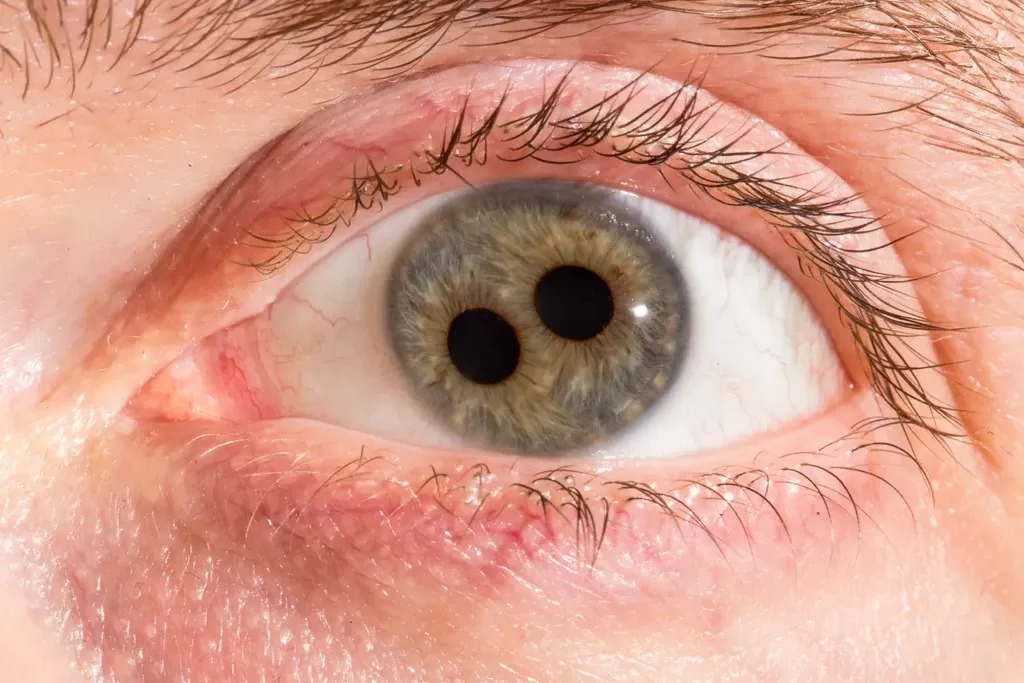
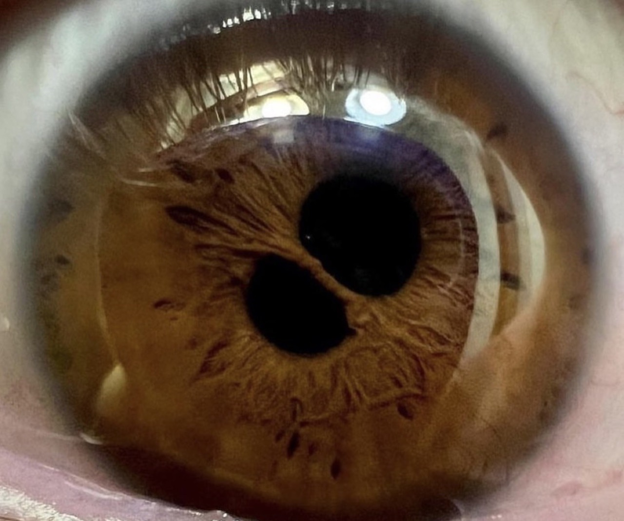
Polycoria means more than one pupillary opening in a single iris.
Most cases seen in clinic are false, with holes that lack a sphincter muscle.
True polycoria, in contrast, is so rare that some authors doubt it exists.
Therefore, the first task is to decide which type is present.
What Polycoria Means
The term combines the Greek words for “many” and “pupil.”
Each opening lets light reach the retina, so optical symptoms may follow.
However, only a functional pupil has its own sphincter and reacts to light.
As a result, the light reflex separates true from false openings.
True Polycoria
True polycoria requires two or more separate pupils, each with an intact sphincter.
Each opening constricts and dilates on its own.
The defect is congenital and usually sporadic.
Also, many older reports may have described simple iris holes.
All-fit Slit-Lamp Adapter
Record and share exactly what you see at the slit lamp. One adapter fits any slit lamp or surgical microscope — and any smartphone.
From Choroida — the team behind this sitePseudopolycoria: The Common Form
Pseudopolycoria describes extra iris holes without sphincter tissue.
These openings stay fixed when a bright light falls on the eye.
Meanwhile, the main pupil constricts normally.
Because of this, the sign points to stromal loss, membrane traction or previous surgery.
Causes of Pseudopolycoria
- Iridocorneal endothelial (ICE) syndrome, especially essential iris atrophy
- Axenfeld-Rieger syndrome with stromal hypoplasia and corectopia
- Persistent pupillary membrane strands that divide the pupil
- Peripheral iridectomy or laser iridotomy
- Blunt trauma with iridodialysis
- Ischemic iris atrophy after acute angle closure or herpes zoster
ICE Syndrome and Polycoria
Essential iris atrophy is the classic acquired cause in adults.
Abnormal corneal endothelium migrates across the angle and iris surface.
Then the membrane contracts, pulling the pupil and tearing new stromal holes.
Therefore, a unilateral case in a middle-aged woman needs specular microscopy and gonioscopy.
Secondary glaucoma usually drives the long-term prognosis.
Axenfeld-Rieger Syndrome
This dysgenesis usually affects both eyes.
The examiner sees posterior embryotoxon, iris strands to Schwalbe line, and stromal thinning.
In addition, dental anomalies and redundant periumbilical skin suggest the systemic form.
About half of patients develop glaucoma, so pressure checks continue for life.
Related dysgeneses also deserve review (see Peters anomaly).
Slit Lamp Examination of Polycoria
First, examine each opening in dim light and then with a bright beam.
A true pupil shows a sphincter ring and constricts briskly.
Next, inspect the surrounding stroma for thinning, ectropion uveae or synechiae.
Retroillumination also reveals transillumination defects beyond the visible holes.
Gonioscopy and Pressure
Gonioscopy is mandatory in every case of pseudopolycoria.
High peripheral anterior synechiae point toward ICE syndrome.
Prominent iris processes to an anterior Schwalbe line favor Axenfeld-Rieger syndrome.
Finally, record baseline pressure and optic disc appearance.
Visual Impact of Polycoria
Many patients have normal acuity and no complaints.
However, extra openings can cause glare, photophobia and monocular diplopia.
Symptoms depend on hole size and position relative to the visual axis.
Large openings may also reduce image quality in bright light.
Polycoria in Children
In a child, the main concern is amblyopia.
Pupil distortion, refractive error or glaucoma may disturb visual development.
Therefore, perform cycloplegic refraction and treat anisometropia early.
Differential Diagnosis of Polycoria
Corectopia describes a single displaced pupil without extra openings.
An iris coloboma forms a keyhole defect continuous with the pupil.
Posterior synechiae distort the pupil outline after uveitis.
Also, an intermittent peaked pupil suggests dilator spasm (see tadpole pupil).
Imaging and Documentation
Slit lamp photography records the number and size of holes.
Anterior segment OCT shows stromal thickness and angle anatomy.
Then, serial images reveal progression, which favors an acquired process.
Managing Polycoria Symptoms
Asymptomatic patients need observation only.
For glare, an opaque iris print contact lens can mask the extra holes.
However, contact lenses need careful hygiene and may not suit young children.
Sunglasses and a brimmed hat help in bright light.
Surgical Repair in Polycoria
Pupilloplasty closes holes with iris sutures, such as McCannel or Siepser techniques.
Surgeons often combine repair with cataract surgery.
However, thin atrophic stroma may not hold sutures well.
In that case, an artificial iris implant suits selected eyes.
Surgery cannot treat the underlying glaucoma or endothelial disease.
Counseling Patients About Polycoria
Explain that the extra opening is usually a simple iris hole without its own muscle.
Then, describe the cause and whether it can progress.
Also, stress regular pressure checks in ICE and Axenfeld-Rieger syndromes.
Affected families benefit from genetic counseling because inheritance is autosomal dominant.
Pitfalls to Avoid
- Do not call an iris hole true polycoria before testing its light reaction
- Never skip gonioscopy and pressure measurement
- Consider ICE syndrome in any unilateral adult case
- Watch for amblyopia in children
- Do not promise better vision after pupilloplasty


Document what you see
Two smartphone imaging tools built for everyday clinic use — one for the slit lamp, one for the fundus.
From Choroida — the team behind this siteReferences
- Idrees F, Vaideanu D, Fraser SG, Sowden JC, Khaw PT. A review of anterior segment dysgeneses. Surv Ophthalmol. 2006;51:213-231.
- Shields MB, Buckley E, Klintworth GK, Thresher R. Axenfeld-Rieger syndrome: a spectrum of developmental disorders. Surv Ophthalmol. 1985;29:387-409.
- Sacchetti M, Mantelli F, Marenco M, et al. Diagnosis and management of iridocorneal endothelial syndrome. Biomed Res Int. 2015;2015:763093.
- Laganowski HC, Kerr Muir MG, Hitchings RA. Glaucoma and the iridocorneal endothelial syndrome. Arch Ophthalmol. 1992;110:346-350.
- Bowling B. Kanski’s Clinical Ophthalmology: A Systematic Approach. 8th ed. Elsevier; 2016.
- American Academy of Ophthalmology. Basic and Clinical Science Course, Section 6: Pediatric Ophthalmology and Strabismus. San Francisco: AAO.
Test yourself
A few questions straight from this article.
-
Which finding defines true polycoria?
True polycoria requires two or more separate pupils, each with its own intact sphincter that constricts and dilates independently; it is so rare that some authors doubt it exists. -
Which clinical test separates true polycoria from pseudopolycoria?
Only a functional pupil has a sphincter and reacts to light; pseudopolycoria holes stay fixed under a bright beam while the main pupil constricts. -
What is the classic acquired cause of pseudopolycoria in adults?
Essential iris atrophy, part of the iridocorneal endothelial syndrome, is the classic acquired adult cause, so any unilateral adult case should raise ICE syndrome. -
How does iridocorneal endothelial (ICE) syndrome create new iris holes?
Abnormal corneal endothelium migrates across the angle and iris surface; the membrane then contracts, pulling the pupil and tearing new stromal holes. -
In pseudopolycoria, which gonioscopic finding favors Axenfeld-Rieger syndrome over ICE syndrome?
Gonioscopy is mandatory in pseudopolycoria: prominent iris processes to an anterior Schwalbe line favor Axenfeld-Rieger syndrome, whereas high peripheral anterior synechiae point to ICE syndrome. -
Roughly what proportion of patients with Axenfeld-Rieger syndrome develop glaucoma?
About half of patients with Axenfeld-Rieger syndrome develop glaucoma, so pressure checks continue for life. -
Which extraocular findings suggest the systemic form of Axenfeld-Rieger syndrome?
Alongside posterior embryotoxon, iris strands to Schwalbe line and stromal thinning, dental anomalies and redundant periumbilical skin suggest the systemic form. -
What is the main visual concern in a child with polycoria?
Pupil distortion, refractive error or glaucoma can disturb visual development, so children need cycloplegic refraction and early treatment of anisometropia. -
Which polycoria mimic forms a keyhole iris defect continuous with the pupil?
An iris coloboma forms a keyhole defect continuous with the pupil, whereas corectopia is a single displaced pupil with no extra openings. -
Which non-surgical option can mask extra iris holes that cause glare in polycoria?
Asymptomatic patients are observed; for glare an opaque iris print contact lens can mask the holes, though lenses need careful hygiene and may not suit young children.

Writes and reviews the clinical articles on Educate. Every post is checked against peer-reviewed sources before it goes live.