Multiple myeloma is a plasma cell cancer that can affect the eye in several ways.
The most important retinal change is hyperviscosity retinopathy.
Therefore, an unexplained dilated retinal vein in an older adult should prompt a blood test for paraprotein.
What Multiple Myeloma Is
Malignant plasma cells fill the bone marrow and produce a monoclonal protein.
This protein can damage bone, kidneys and the blood.
Also, the International Myeloma Working Group sets diagnostic criteria.
As a result, a clear definition guides testing.
Eye Findings in Multiple Myeloma
Knapp and colleagues described ocular findings in myeloma.
They include retinopathy, ciliary body cysts and corneal deposits.
Also, orbital plasmacytoma can cause proptosis.
Therefore, the eye can show many signs.
Fundus Explorer Pro
Photograph the retinal findings described here with the phone already in your pocket — 22 D optics and built-in illumination in one handheld unit.
From Choroida — the team behind this siteHyperviscosity Syndrome
High paraprotein levels thicken the blood.
Flow slows in small vessels, and symptoms follow.
Also, hyperviscosity is more common in IgM disease and in some IgA myeloma.
As a result, eye signs can appear before other symptoms.
Fundus Findings in Multiple Myeloma Retinopathy
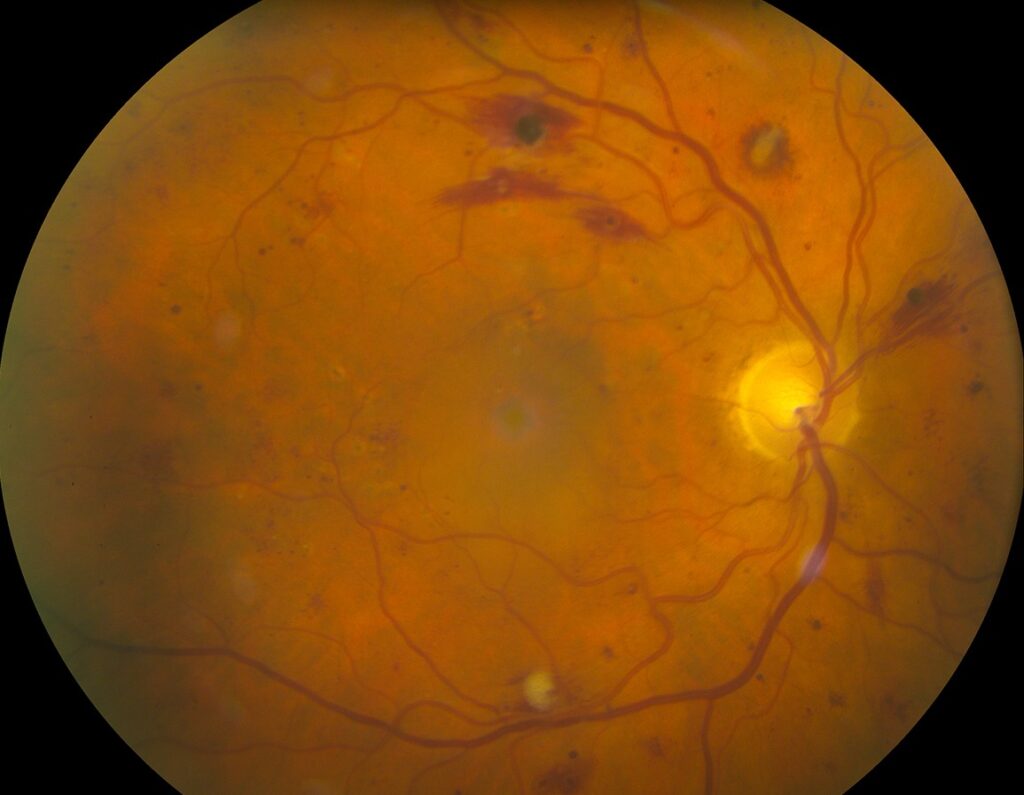
Veins become dilated and tortuous with sausage-like segments.
Hemorrhages occur in all layers of the retina.
Also, microaneurysms and cotton-wool spots appear.
In addition, the disc may swell.
Comparison with Vein Occlusion
Hyperviscosity retinopathy is bilateral and diffuse.
Central vein occlusion is usually unilateral and more severe in the affected eye.
Also, venous dilation in both eyes suggests systemic disease (see central retinal vein occlusion).
Therefore, bilateral findings need a blood workup.
Waldenström Macroglobulinemia
Menke and colleagues described hyperviscosity retinopathy in Waldenström macroglobulinemia.
The retinal findings correlate with serum viscosity.
Also, plasma exchange reverses the changes.
As a result, the same lessons apply to myeloma.
Mechanisms of Multiple Myeloma Retinopathy
Large proteins increase blood viscosity and cause stasis.
Endothelial damage and hypoxia lead to leakage.
Also, paraprotein can deposit in the vessel wall.
Therefore, ischemia and bleeding follow.
Other Ocular Manifestations
Crystalline keratopathy occurs from light chain deposits.
Ciliary body cysts and glaucoma are rare.
Also, optic nerve infiltration is possible.
In addition, drug toxicity can affect the eye.
Differential Diagnosis
Diabetic retinopathy, hypertension and leukemia cause hemorrhages.
Polycythemia and sickle cell disease cause venous changes.
Also, anemia produces hemorrhages with pale discs.
Therefore, use blood tests to separate the causes.
Investigations in Multiple Myeloma
Order serum protein electrophoresis, immunofixation and free light chains.
Measure serum viscosity and a complete blood count.
Also, check calcium, kidney function and imaging of bone.
As a result, the diagnosis is confirmed.
Ocular Examination
Perform a dilated fundus exam and OCT.
Fluorescein angiography shows slow filling and leakage.
Also, record the findings with photographs.
In addition, repeat after treatment.
Treatment of Hyperviscosity
Plasma exchange lowers viscosity quickly.
It is used for symptomatic hyperviscosity.
Also, systemic therapy reduces the paraprotein over time.
Therefore, urgent referral to a hematologist is needed.
Systemic Treatment of Myeloma
Modern therapy includes proteasome inhibitors, immunomodulatory drugs and monoclonal antibodies.
Autologous transplant is an option in fit patients.
Also, Kyle and Rajkumar reviewed these strategies.
As a result, survival has improved.
Ocular Side Effects of Treatment
Some drugs cause dry eye, keratopathy or blurred vision.
Antibody-drug conjugates are linked to corneal toxicity.
Also, steroids raise the risk of cataract and glaucoma.
In addition, monitor patients with eye symptoms.
Prognosis in Multiple Myeloma Retinopathy
Retinopathy often improves after reduction of viscosity.
Vision loss from hemorrhage or edema may be permanent.
Also, the retinal signs can guide the pace of treatment.
Therefore, early recognition helps.
Pitfalls to Avoid
- Do not label bilateral venous dilation as diabetic without a blood test
- Never delay referral in suspected hyperviscosity
- Do not forget to review drug effects on the eye
- Never stop eye follow-up once the viscosity falls
Monoclonal Gammopathy of Undetermined Significance
Many older patients have a monoclonal protein without myeloma.
They rarely develop hyperviscosity.
Also, eye findings are uncommon in this group.
Therefore, a mild paraprotein seldom explains retinopathy.
Light Chain Deposition in the Eye
Light chains can deposit in the cornea, ciliary body and conjunctiva.
They can cause crystalline changes and pain.
Also, some patients show a Fanconi syndrome.
As a result, the cornea can offer a diagnostic clue.
Orbital and Optic Nerve Involvement
Plasmacytomas can invade the orbit and cause proptosis and diplopia.
Optic nerve compression is possible.
Also, MRI shows a mass in the bone or orbit.
Therefore, image the orbit in suspected cases.
Role of the Ophthalmologist
The eye specialist may be first to suspect disease.
A prompt call to the hematologist can start therapy.
Also, a baseline eye exam helps later monitoring.
In addition, document the findings carefully.
For the latest studies, see the PubMed results for multiple myeloma.


Document what you see
Two smartphone imaging tools built for everyday clinic use — one for the slit lamp, one for the fundus.
From Choroida — the team behind this siteReferences
- Knapp AJ, Gartner S, Henkind P. Multiple myeloma and its ocular manifestations. Surv Ophthalmol. 1987;31:343-351.
- Menke MN, Feke GT, McMeel JW, Branagan A, Hunter Z, Treon SP. Hyperviscosity-related retinopathy in Waldenström macroglobulinemia. Arch Ophthalmol. 2006;124:1601-1606.
- Rajkumar SV, Dimopoulos MA, Palumbo A, et al. International Myeloma Working Group updated criteria for the diagnosis of multiple myeloma. Lancet Oncol. 2014;15:e538-e548.
- Kyle RA, Rajkumar SV. Multiple myeloma. Blood. 2008;111:2962-2972.

Writes and reviews the clinical articles on Educate. Every post is checked against peer-reviewed sources before it goes live.