Vitreous biopsy collects vitreous fluid for laboratory tests when the cause of vitritis or retinal disease remains unclear.
It helps to diagnose infection, intraocular lymphoma, and other masquerade syndromes.
The yield depends on careful planning, timing, and communication with the laboratory.
Indications for Vitreous Biopsy
- Suspected endophthalmitis, either endogenous or postoperative
- Posterior uveitis that does not respond to treatment
- Suspected vitreoretinal lymphoma
- Atypical retinitis, such as necrotizing herpetic retinitis or toxoplasmosis
Consider a biopsy when noninvasive tests fail to give a diagnosis and the result will change management (see uveitis masquerade syndromes).
Preparing for Vitreous Biopsy
Discuss the plan with the pathologist and microbiologist before surgery.
Ask which tests to prioritize, since the sample is small.
Avoid corticosteroids before the biopsy when possible, because steroids reduce the yield of cytology in lymphoma.
Plan to transport the sample immediately, since delay degrades cells.
Fundus Explorer Pro
Photograph the retinal findings described here with the phone already in your pocket — 22 D optics and built-in illumination in one handheld unit.
From Choroida — the team behind this siteVitreous Biopsy by Needle or Vitrectomy
A needle tap uses a 23 to 25-gauge needle on a syringe to aspirate 0.2 to 0.5 mL, and it suits suspected endophthalmitis.
A vitrectomy biopsy uses a cutter with the infusion line off, so the sample remains undiluted.
Surgeons collect 0.5 to 1 mL, then turn the infusion on and perform the planned surgery.
Vitrectomy gives a larger and cleaner sample, while a needle tap carries a lower surgical burden (see pars plana vitrectomy).
Tests to Order After Vitreous Biopsy
Cytology and flow cytometry look for lymphoma cells, and immunohistochemistry adds further detail.
The ratio of interleukin 10 to interleukin 6 helps with lymphoma, since a higher interleukin 10 level favors it.
Polymerase chain reaction detects herpesviruses, toxoplasma, and bacterial or fungal DNA.
Cultures identify bacteria and fungi and give antibiotic sensitivities.
Interpreting Vitreous Biopsy Results
A negative cytology does not exclude lymphoma, since the cells are fragile and sparse.
Repeat the biopsy or combine it with chorioretinal biopsy when suspicion remains high.
A positive PCR confirms infection, but a negative result does not exclude it (see intraocular lymphoma management).
Complications of Vitreous Biopsy
- Retinal tear or detachment
- Vitreous hemorrhage
- Endophthalmitis, which is rare
- Hypotony from leaking ports
Surgeons lower the risk with good wound closure and careful peripheral examination.
An Example Case
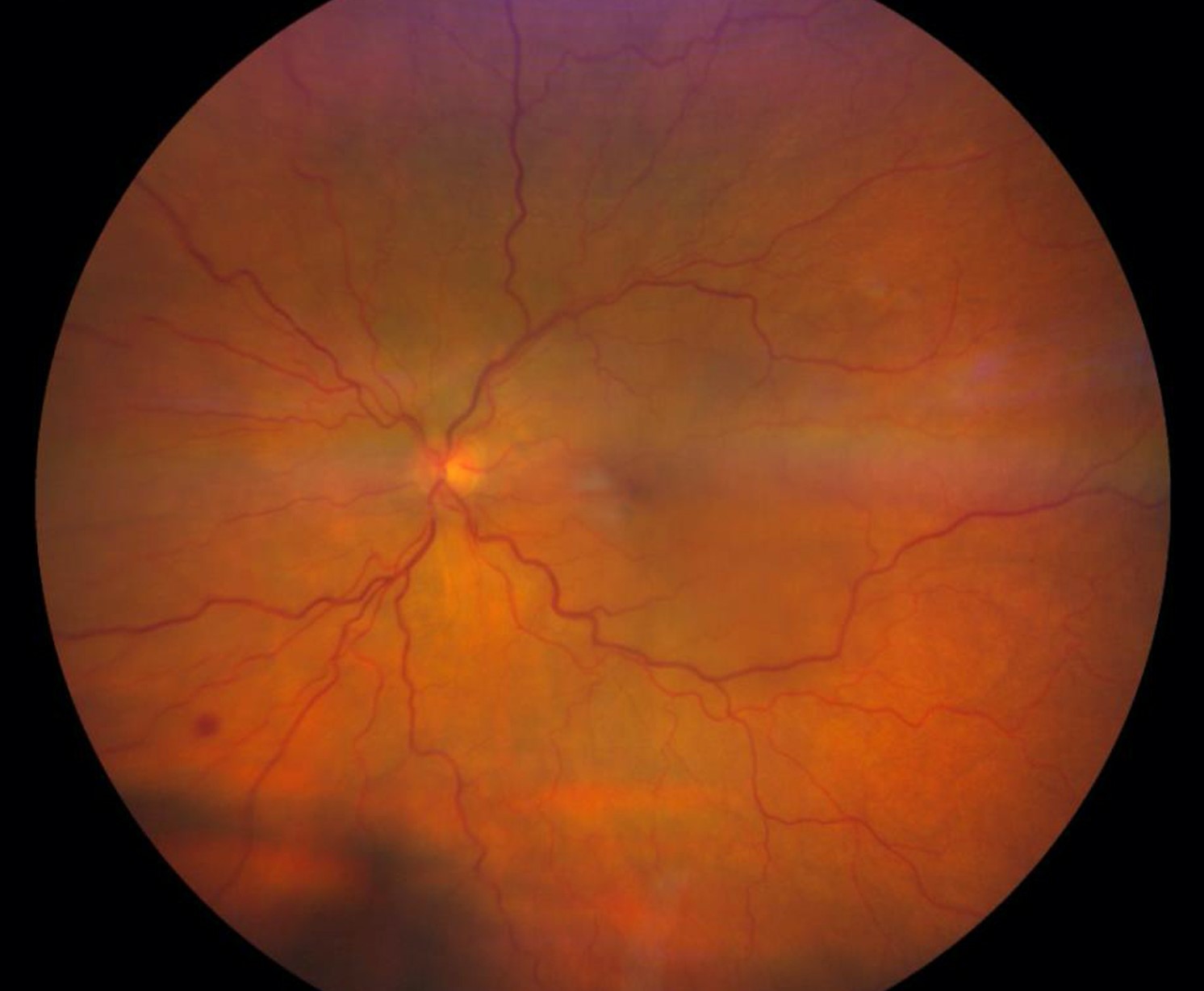
A 68-year-old man has chronic vitritis and subretinal lesions that did not respond to steroids.
After a washout period, he undergoes a diagnostic vitrectomy with an undiluted sample.
Cytology shows large atypical B cells, and the interleukin ratio supports lymphoma.
Imaging of the brain finds no lesions, and the oncology team starts therapy.
Key Points
- Collect an undiluted sample first
- Coordinate with the laboratory in advance
- Withhold steroids before biopsy when safe
Questions Patients Ask
Patients ask whether the biopsy is painful, and the surgery uses local or general anesthesia.
They also ask when results arrive, and cultures take days while cytology takes a few days.
Because the result may change treatment, explain that the surgeon may begin therapy before all tests finish.
Pitfalls to Avoid
- Giving steroids before biopsy in suspected lymphoma
- Diluting the first sample with infusion fluid
- Sending the sample late, after cells have degraded
- Relying on one negative test to exclude lymphoma
Looking Ahead
Next-generation sequencing of vitreous fluid is entering practice, and it finds rare pathogens.
Also, liquid biopsy of aqueous humor may reduce the need for vitrectomy in some cases.
However, vitreous biopsy stays the main route when the diagnosis is uncertain.
Practical Points
Label the tubes before the case, and tell the laboratory the suspected diagnosis.
Then prepare the transport medium and keep the sample cool when instructed.
Also document the volume and the time of collection.
Handling the Specimen
Divide the sample according to the planned tests, and prioritize cytology and PCR when the volume is small.
Also transport cytology in a fixative that the laboratory recommends.
Then send the culture sample in a sterile container without delay.
Because every minute counts for cell survival, call the laboratory before you leave the operating room.
Safety After Biopsy
Examine the eye the day after surgery for pressure, inflammation, and retinal breaks.
Then arrange a visit at one week to review results and plan treatment.
Because the biopsy may change the diagnosis, communicate with the uveitis or oncology team early.
Also document the exact tests sent in the chart, so that results can be traced.


Document what you see
Two smartphone imaging tools built for everyday clinic use — one for the slit lamp, one for the fundus.
From Choroida — the team behind this siteReferences
- Davis JL, Miller DM, Ruiz P. Diagnostic testing of vitrectomy specimens. Am J Ophthalmol. 2005;140:822-829.
- Whitcup SM, De Smet MD, Rubin BI, et al. Intraocular lymphoma: clinical and histopathologic diagnosis. Ophthalmology. 1993;100:1399-1406.
- Chan CC, Rubenstein JL, Coupland SE, et al. Primary vitreoretinal lymphoma: a report from an International Primary Central Nervous System Lymphoma Collaborative Group symposium. Oncologist. 2011;16:1589-1599.
- American Academy of Ophthalmology. Basic and Clinical Science Course, Section 12: Retina and Vitreous. AAO; current edition.

Writes and reviews the clinical articles on Educate. Every post is checked against peer-reviewed sources before it goes live.