Central scotoma is a blind or dim area at fixation, and it reduces reading and face recognition even when peripheral vision is normal.
It arises from diseases of the macula, the optic nerve, or less often the chiasm and visual pathway.
A structured approach separates these causes quickly and avoids unnecessary imaging.
What Central Scotoma Means
The macula and papillomacular bundle serve the central 10 degrees of the visual field.
Damage anywhere along this route produces a central or centrocecal defect.
The size and density of the scotoma help to guide the clinician toward the cause.
Causes of Central Scotoma
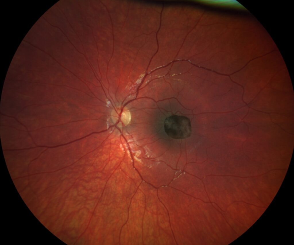
- Macular disease: AMD, central serous disease, macular hole, dystrophies
- Optic neuropathy: optic neuritis, compression, toxic and nutritional causes
- Hereditary disease: Leber hereditary optic neuropathy and dominant optic atrophy
- Occipital pole lesions and other pathway disease
Walsh and Hoyt describe these differences in detail in their neuro-ophthalmology text (see toxic nutritional optic neuropathy).
Fundus Explorer Pro
Photograph the retinal findings described here with the phone already in your pocket — 22 D optics and built-in illumination in one handheld unit.
From Choroida — the team behind this siteExamination in Central Scotoma
Measure acuity, pinhole acuity, and color vision, and check pupil reactions for a relative afferent defect.
Perform an Amsler grid test, and map the scotoma with a tangent screen or a 10-2 field.
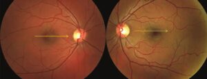
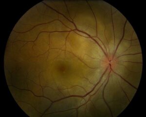
Dilate the pupil and inspect the macula and optic disc carefully.
Because a normal fundus does not exclude disease, add OCT and consider ERG when the cause remains unclear.
Macular Versus Optic Nerve Causes
Macular disease usually causes metamorphopsia and a positive photostress test.
Optic nerve disease causes marked color desaturation, a relative afferent defect, and a field defect that respects the nerve fiber pattern.
However, OCT of the macula and nerve fiber layer helps in doubtful cases (see Leber hereditary optic neuropathy).
Imaging and Tests in Central Scotoma
- OCT of the macula and the ganglion cell layer
- Fundus autofluorescence for RPE disease
- Visual evoked potentials and pattern ERG when the cause is unclear
- MRI of the orbits and brain with contrast when the optic nerve is suspected
Then order tests in a sequence that follows the clinical findings.
Management
When treatment exists, such as anti-VEGF for neovascular AMD or steroids for optic neuritis, treat the cause.
Also refer patients with permanent scotomas for low vision rehabilitation and training of eccentric viewing.
An Example Case
A 32-year-old man notices a blur in the center of the left eye over two weeks, and acuity is 20/100.
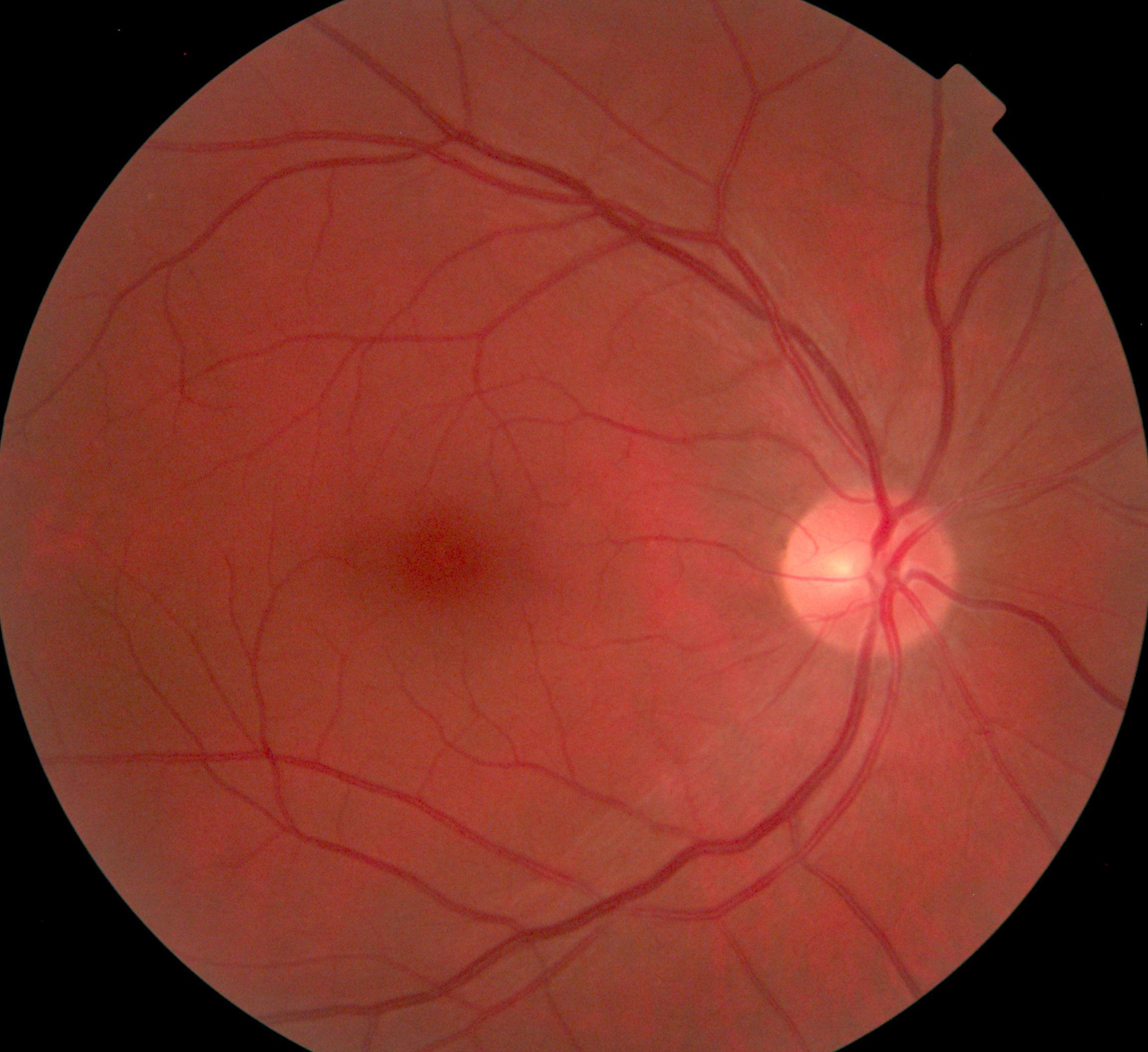
The fundus looks normal, and there is a relative afferent pupillary defect and marked color desaturation.
On the 10-2 field, a dense central scotoma appears, and MRI shows enhancement of the optic nerve.
So the diagnosis is optic neuritis, and he receives treatment and recovers to 20/25.
A normal macula on OCT and an abnormal color test pointed to the nerve from the start.
Questions Patients Ask
Patients ask whether the scotoma will fill in, and the answer depends on the cause.
Some causes, such as optic neuritis, often improve, while others, such as geographic atrophy, do not.
Because the scotoma affects reading, offer practical advice on large print and good lighting.
Pitfalls to Avoid
- Assuming that a normal fundus excludes macular disease
- Ordering MRI before checking pupils and color vision
- Ignoring a central scotoma in a patient taking ethambutol or linezolid
- Forgetting the possibility of nonorganic visual loss when tests are normal
Looking Ahead
Better imaging of the ellipsoid zone and the ganglion cell layer helps to sort causes earlier.
Also, genetic testing is becoming standard in young patients with unexplained scotomas.
Therefore, the workup may become more targeted.
Practical Points
Record the size and the density of the scotoma on the chart with an Amsler grid drawing.
Then repeat the field after treatment to document change.
Also check drug history in every patient with a bilateral central scotoma.
Eccentric Viewing With a Central Scotoma
Patients with a dense scotoma learn to use a preferred retinal locus away from the fovea.
Low vision services can train this skill and improve reading speed.
Because the adjustment takes weeks, refer early.
Documenting the Scotoma
Draw the scotoma on a grid or record it with a 10-2 field.
Then compare size and density at each visit, since change guides treatment.
Because patients often fill in missing areas, ask them to point at the blank region with a pen.
Also record whether the scotoma touches fixation, since this predicts reading difficulty.
Associated Symptoms
Photophobia and hemeralopia suggest cone disease.
Pain on eye movement suggests optic neuritis.
Because systemic symptoms may point to the cause, review neurologic and toxic exposures.


Document what you see
Two smartphone imaging tools built for everyday clinic use — one for the slit lamp, one for the fundus.
From Choroida — the team behind this siteReferences
- Miller NR, Newman NJ, Biousse V, Kerrison JB, eds. Walsh and Hoyt’s Clinical Neuro-Ophthalmology. 6th ed. Lippincott Williams & Wilkins; 2005.
- Trobe JD. The Neurology of Vision. Oxford University Press; 2001.
- Gass JDM. Stereoscopic Atlas of Macular Diseases: Diagnosis and Treatment. 4th ed. Mosby; 1997.
- American Academy of Ophthalmology. Basic and Clinical Science Course, Section 5: Neuro-Ophthalmology. AAO; current edition.

Writes and reviews the clinical articles on Educate. Every post is checked against peer-reviewed sources before it goes live.