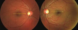
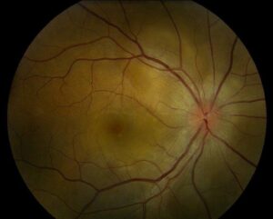
AV nicking is the apparent narrowing of a retinal vein where an artery crosses it.
It results from thickened arteriolar walls that press on the vein.
Therefore, it is a sign of chronic hypertension and small vessel disease.
Anatomy of the Crossing
Retinal arteries and veins share a common adventitial sheath at crossings.
The artery usually lies over the vein.
Also, the two vessels cannot slide apart.
As a result, a thick artery presses directly on the vein.
What AV Nicking Looks Like
The vein tapers on both sides of the crossing.
It may seem to disappear under the artery.
Also, the vein often appears dilated beyond the crossing.
Therefore, the crossing looks like a pinch.
Fundus Explorer Pro
Photograph the retinal findings described here with the phone already in your pocket — 22 D optics and built-in illumination in one handheld unit.
From Choroida — the team behind this siteNamed Signs at the Crossing
Gunn’s sign is the tapering of the vein on each side of the crossing.
Salus’ sign is a deflection of the vein at the crossing in an S shape.
Also, Bonnet’s sign is banking of the vein distal to the crossing.
In addition, these signs track increasing severity.
Why AV Nicking Occurs
Long-standing hypertension thickens and hardens the arteriolar wall.
The process is called arteriolosclerosis.
Also, the rigid artery compresses the vein with each pulse.
As a result, flow in the vein slows and the vessel narrows.
AV Nicking and Hypertension
Klein and colleagues found AV nicking in a population-based study and linked it to long-term blood pressure.
The sign reflects chronic and not acute elevation.
Also, it persists after pressure control.
Therefore, it marks past exposure and not current levels (see hypertensive retinopathy).
Related Signs of Arteriolosclerosis
Arteriolar wall thickening produces copper and silver wiring.
Focal narrowing and widened light reflexes accompany it.
Also, vessel tortuosity can appear.
In addition, these signs often come together.
Grading AV Nicking
Classic grading divides nicking into mild, moderate and severe.
Mild shows slight tapering, and severe shows the vein hidden under the artery.
Also, population studies use photographs and standard grades.
Therefore, grading needs good images and consistent rules.
Risk of Stroke and Heart Disease
Wong and colleagues showed that retinal arteriolar narrowing predicts coronary disease.
AV nicking also relates to stroke risk in large cohorts.
Also, Yatsuya and colleagues linked retinal microvascular signs to lacunar stroke.
As a result, the retina offers a view of the cerebral small vessels.
Hypertension Risk Prediction
Retinal arteriolar narrowing predicts the later development of hypertension.
Wong and colleagues found this in a large cohort.
Also, the effect held after adjusting for other risk factors.
Therefore, early narrowing may signal a risk before the diagnosis.
AV Nicking and Retinal Vein Occlusion
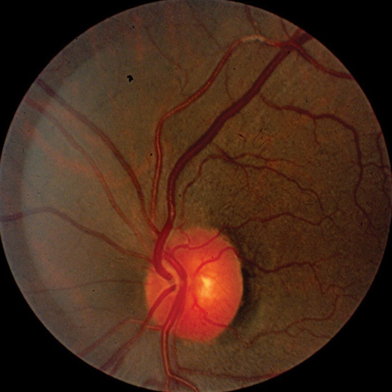
Branch retinal vein occlusion usually occurs at an arteriovenous crossing.
The shared sheath compresses the vein and promotes thrombosis (see branch retinal vein occlusion).
Also, hypertension is the main risk factor.
As a result, AV nicking warns of this complication.
Differential Diagnosis
Normal crossings can look narrow in some eyes.
Vein compression can also occur with tumors or scars.
Also, tilted discs alter vessel paths.
Therefore, look for the whole pattern and not a single crossing.
Examination Tips for AV Nicking
Use a dilated examination or a good photograph.
Look at crossings near the disc where vessels are large.
Also, compare both eyes.
In addition, record the number of crossings affected.
Imaging Options
Color photographs allow grading in a standard way.
Red-free images show vessel borders clearly.
Also, OCT angiography can show reduced vessel density.
As a result, imaging supports risk assessment.
Managing a Patient With AV Nicking
Treat the underlying hypertension and other vascular risk factors.
Refer the patient for a full cardiovascular review.
Also, check kidney function and lipids.
Therefore, the retinal sign triggers a systemic assessment.
Counseling Patients
Explain that the sign shows long-term stress on the small vessels.
Say that control of blood pressure reduces further damage.
Also, recommend regular checks.
In addition, give clear advice on diet and exercise.
Pitfalls to Avoid
- Do not call every tapered vein AV nicking without seeing the crossing pattern
- Never treat the sign as acute hypertension
- Do not forget to refer the patient for systemic care
- Never rely on a single photograph for grading
Measuring Retinal Vessel Caliber
Software can measure vessel width from photographs.
Results are summarized as central retinal arteriolar and venular equivalents.
Also, the arteriole-to-venule ratio falls with narrowing.
Therefore, these tools support research and risk scoring.
Children and Young Adults
Young patients with severe hypertension can show arteriolar narrowing without nicking.
Crossing changes need years of vessel wall thickening.
Also, the absence of nicking does not exclude hypertension.
Therefore, read the sign with age in mind.


Document what you see
Two smartphone imaging tools built for everyday clinic use — one for the slit lamp, one for the fundus.
From Choroida — the team behind this siteReferences
- Wong TY, Mitchell P. Hypertensive retinopathy. N Engl J Med. 2004;351:2310-2317.
- Klein R, Klein BEK, Moss SE, Wang Q. Hypertension and retinopathy, arteriolar narrowing, and arteriovenous nicking in a population. Arch Ophthalmol. 1994;112:92-98.
- Wong TY, Klein R, Sharrett AR, et al. Retinal arteriolar narrowing and risk of coronary heart disease in men and women: the Atherosclerosis Risk in Communities Study. JAMA. 2002;287:1153-1159.
- Wong TY, Klein R, Sharrett AR, et al. Retinal arteriolar diameter and risk for hypertension. Ann Intern Med. 2004;140:248-255.

Writes and reviews the clinical articles on Educate. Every post is checked against peer-reviewed sources before it goes live.