Alzheimer disease and the retina are linked because the retina is a thin extension of the brain.
Researchers look at it for early signs of neurodegeneration.
Therefore, retinal imaging could one day help to find patients at risk.
Why Look at the Retina
The retina is part of the central nervous system and is easy to image.
Its neurons and vessels share features with the brain.
Also, imaging is quick, cheap and noninvasive.
As a result, it has drawn interest as a window into brain disease.
Retinal Nerve Fiber Layer in Alzheimer Disease
Meta-analyzes show thinner retinal nerve fiber layer in Alzheimer disease than in controls.
Chan and colleagues pooled spectral-domain OCT studies and found a consistent effect.
Also, the superior and inferior quadrants show the largest changes.
Therefore, the nerve fiber layer is the best studied marker.
Fundus Explorer Pro
Photograph the retinal findings described here with the phone already in your pocket — 22 D optics and built-in illumination in one handheld unit.
From Choroida — the team behind this siteMacular Thickness in Alzheimer Disease
The ganglion cell layer and inner plexiform layer thin with disease.
Total macular thickness falls in some cohorts.
Also, den Haan and colleagues reviewed these findings across many studies.
As a result, inner retinal measures show promise.
Mild Cognitive Impairment
Patients with mild cognitive impairment show intermediate thinning.
This suggests that retinal changes begin early.
Also, some studies link thinning to later conversion to dementia.
However, the evidence is not yet strong enough for clinical use.
Amyloid in the Retina of Alzheimer Disease
Koronyo-Hamaoui and colleagues reported amyloid-beta deposits in the retina of patients with Alzheimer disease.
The deposits lie in the inner layers and near vessels.
Also, they correlate with brain pathology in some studies.
Therefore, the retina may mirror the brain.
Imaging Amyloid in Living Eyes
Koronyo and colleagues used curcumin fluorescence to image retinal amyloid in patients.
The signal was higher in patients with the disease.
Also, the method is still experimental.
As a result, it needs confirmation in larger groups.
Hyperspectral Imaging
Hadoux and colleagues used hyperspectral imaging to detect spectral changes in the retina linked to brain amyloid.
The test distinguished patients with positive brain scans.
Also, it needs no dye.
Therefore, it might serve as a screening tool in the future.
Retinal Vascular Changes in Alzheimer Disease
OCT angiography shows reduced vessel density in patients with Alzheimer disease.
The foveal avascular zone may enlarge.
Also, retinal venules can narrow.
As a result, the microvascular changes echo those of the brain.
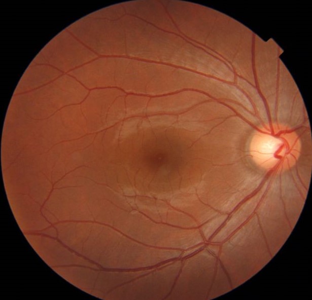
Functional Changes in Alzheimer Disease
Contrast sensitivity and color vision decline in some patients.
Electroretinography can show changes in the inner retina.
Also, visual field defects occur.
In addition, pupil responses may be abnormal.
Visual Symptoms in Dementia
Patients may complain of difficulty reading or recognizing faces.
These problems come mostly from the brain and not from the eye.
Also, ocular disease adds to the burden.
Therefore, test eyes regularly in patients with dementia (see glaucoma and the optic disc).
Alzheimer Disease and the Retina in Other Conditions
Glaucoma, age-related macular degeneration and diabetes also thin the inner retina.
These diseases can confuse the picture.
Also, Parkinson disease and multiple sclerosis show similar patterns.
As a result, retinal thinning is not specific.
Why OCT Is Not Yet a Diagnostic Test
Measurements overlap between patients and healthy people.
Devices and software differ, and normal ranges vary.
Also, eye disease and age affect the numbers.
Therefore, OCT cannot diagnose or exclude the disease.
Ongoing Research
Studies combine OCT, angiography and artificial intelligence.
Large cohorts such as the UK Biobank help to find patterns.
Also, trials aim to link retinal change to biomarkers in the blood and spinal fluid.
In addition, longer follow-up will clarify the predictive value.
What Clinicians Can Do Now
Perform routine eye care in patients with cognitive decline.
Refer patients with new visual complaints for a full eye examination.
Also, treat cataract and glaucoma, which can worsen function.
As a result, quality of life improves.
Counseling Patients
Explain that retinal tests are research tools.
Say that an abnormal scan does not mean dementia.
Also, avoid sending patients to commercial screening that promise diagnosis.
Therefore, keep the message clear and honest.
Pitfalls to Avoid
- Do not tell a patient that a thin nerve fiber layer means Alzheimer disease
- Never skip a search for glaucoma in a patient with thinning
- Do not rely on retinal tests for diagnosis
- Never forget that cataract and eye disease reduce function in dementia
For the latest studies, see the PubMed results for Alzheimer disease.


Document what you see
Two smartphone imaging tools built for everyday clinic use — one for the slit lamp, one for the fundus.
From Choroida — the team behind this siteReferences
- London A, Benhar I, Schwartz M. The retina as a window to the brain: from eye research to CNS disorders. Nat Rev Neurol. 2013;9:44-53.
- Chan VTT, Sun Z, Tang S, et al. Spectral-domain OCT measurements in Alzheimer’s disease: a systematic review and meta-analysis. Ophthalmology. 2019;126:497-510.
- Koronyo Y, Biggs D, Barron E, et al. Retinal amyloid pathology and proof-of-concept imaging trial in Alzheimer’s disease. JCI Insight. 2017;2:e93621.
- Hadoux X, Hui F, Lim JKH, et al. Non-invasive in vivo hyperspectral imaging of the retina for potential biomarker use in Alzheimer’s disease. Nat Commun. 2019;10:4227.

Writes and reviews the clinical articles on Educate. Every post is checked against peer-reviewed sources before it goes live.