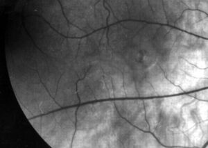
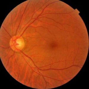
Central retinal artery occlusion in young patients is uncommon, and the causes differ from those in older adults.
Atherosclerosis and carotid disease dominate later in life, while embolic, hematologic, inflammatory, and congenital causes lead in the young.
A thorough workup finds the cause and protects against stroke.
Why Central Retinal Artery Occlusion in Young Patients Is Different
Brown and colleagues described retinal arterial obstruction in children and young adults and found a wide range of causes.
Greven and colleagues reported a similar mix in adults under 40.
Many patients have no typical vascular risk factors, so the clinician must look further (see central retinal artery occlusion management).
Causes of Central Retinal Artery Occlusion in Young Patients
- Cardiac sources: valvular disease, endocarditis, patent foramen ovale, myxoma
- Hypercoagulable states: antiphospholipid syndrome, protein C or S deficiency, factor V Leiden
- Inflammatory disease: vasculitis, Behçet disease, lupus, Susac syndrome
- Migraine, drug use such as cocaine, trauma, and oral contraceptive use
- Sickle cell disease and other hematologic disorders
In some patients, no cause is found.
Fundus Explorer Pro
Photograph the retinal findings described here with the phone already in your pocket — 22 D optics and built-in illumination in one handheld unit.
From Choroida — the team behind this siteExamination in Central Retinal Artery Occlusion in Young Patients
Check visual acuity, pupils, and the fundus for cherry-red spot, emboli, and cilioretinal sparing.
Examine the heart and carotids, and measure blood pressure on both arms.
Look for signs of systemic disease, such as skin lesions and joint pain (see Susac syndrome).
Workup of Central Retinal Artery Occlusion in Young Patients
- ECG, echocardiography with bubble study, and rhythm monitoring
- Carotid and vertebral imaging
- Complete blood count, ESR, CRP, and coagulation studies
- Antiphospholipid antibodies, thrombophilia tests, and autoimmune screening
- Sickle cell testing when relevant
Refer urgently, since an occluded retinal artery may be a stroke equivalent.
Treatment of Central Retinal Artery Occlusion in Young Patients
Treatment of the acute event has limited evidence, and many centers try conservative measures within hours of onset.
Hayreh and Zimmerman reported the visual outcome of conservative and other treatments.
Address the underlying cause, such as anticoagulation for thrombophilia or treatment of the vasculitis.
Prognosis of Central Retinal Artery Occlusion in Young Patients
Young patients may recover better than older ones, especially with cilioretinal sparing.
Long-term care aims to prevent recurrence and stroke.
An Example Case
A 31-year-old woman with migraine and oral contraceptive use has sudden painless loss of vision in the right eye.
Examination shows a cherry-red spot and a cilioretinal artery that preserves a small island of vision.
Echocardiography with a bubble study shows a patent foramen ovale, and no carotid disease.
Thrombophilia tests are negative, and the team stops the contraceptive, starts antiplatelet treatment, and arranges closure of the defect.
Questions Patients Ask
Patients ask whether they will get the vision back, and recovery is limited, though young people do better than older adults.
They also ask whether the other eye is at risk, and the answer depends on the cause.
Because a stroke may follow, explain the need for urgent systemic evaluation.
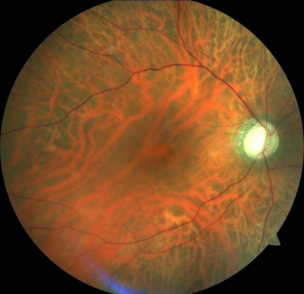
Pitfalls to Avoid
- Attributing the event to age without a full workup
- Skipping echocardiography because the carotid scan is normal
- Missing thrombophilia and autoimmune causes
- Failing to send the patient for a stroke evaluation on the same day
Looking Ahead
Better imaging of cardiac sources and genetic tests for clotting disorders may increase diagnostic yield.
Also, trials of thrombolysis for retinal artery occlusion continue.
Therefore, treatment pathways may change in the next years.
Practical Points
Treat the event as a possible stroke, and use the stroke pathway of your hospital.
Then collect a complete history, including drugs, migraine, and family clotting history.
Also repeat the examination for emboli and neovascularization.
The Role of Cilioretinal Arteries
A cilioretinal artery supplies the papillomacular bundle in many people.
When present, it can preserve central vision during a central occlusion.
Therefore, patients with cilioretinal sparing often have better outcomes.
Follow-Up
Examine at one week, one month, and three months for neovascular glaucoma.
Because ischemia may trigger new vessels, check the angle and the iris.
Then treat with panretinal photocoagulation when needed.
Imaging the Retina in Young Patients
OCT shows inner retinal swelling in the acute phase and later thinning of the inner layers.
Fluorescein angiography shows delayed arterial filling and, in some patients, an embolus.
Because the findings guide the diagnosis, obtain them early.
Also repeat the imaging at follow-up to detect neovascularization.
Counselling About Recurrence
Explain that the risk of a repeat event depends on the cause.
Then outline the plan for prevention, including medications and lifestyle measures.
Because anxiety is common, give a clear contact for new symptoms.


Document what you see
Two smartphone imaging tools built for everyday clinic use — one for the slit lamp, one for the fundus.
From Choroida — the team behind this siteReferences
- Brown GC, Magargal LE, Shields JA, et al. Retinal arterial obstruction in children and young adults. Ophthalmology. 1981;88:18-25.
- Greven CM, Slusher MM, Weaver RG. Retinal arterial occlusions in young adults. Am J Ophthalmol. 1995;120:776-783.
- Hayreh SS, Zimmerman MB. Central retinal artery occlusion: visual outcome. Am J Ophthalmol. 2005;140:376-391.
- American Academy of Ophthalmology. Basic and Clinical Science Course, Section 12: Retina and Vitreous. AAO; current edition.

Writes and reviews the clinical articles on Educate. Every post is checked against peer-reviewed sources before it goes live.