
CASE REPORT
A 35-year-old male, presented to an ophthalmology clinic with a history of blunt trauma to his left eye after being hit by a baseball while playing sports. He experienced decreased visual acuity, persistent ocular pain, and intermittent redness in the affected eye.

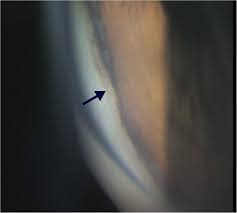
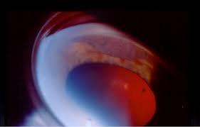
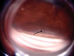
Slit Lamp Examination revealed mild hyperemia in the conjunctiva, a clear cornea, a shallow anterior chamber, mild distortion and atrophy in the inferonasal quadrant of the iris, and no signs of traumatic cataract.
Gonioscopy showed an open angle, while fundus examination revealed a normal optic disc, macula, and peripheral retina. Based on these findings, he was diagnosed with a cyclodialysis cleft in his left eye.
Cyclodialysis Cleft DISEASE entity
A cyclodialysis cleft is a separation of the ciliary body from the scleral spur, creating a direct connection between the anterior chamber and the suprachoroidal space. Many will spontaneously close, but those that do not can cause chronic hypotony, resulting in hypotony maculopathy, optic disc edema, and decreased visual acuity.
Cyclodialysis cleft is formed when the longitudinal fibers of the ciliary muscle separate from the scleral spur, forming a direct connection between the anterior chamber and the suprachoroidal space. This dramatically increases aqueous outflow and predisposes the eye to hypotony.
The most common causes of cyclodialysis cleft are trauma or following various intraocular surgeries including trabeculectomy, trabeculotomy, goniotomy, phacoemulsification extracapsular cataract extraction, secondary intraocular lens (IOL) placement, phakic IOL removal, and displacement of an anterior-chamber IOL.
In modern surgical practice, the creation of a cyclodialysis cleft is rarely intentional; it was once, however, an accepted intervention for open-angle and aphakic glaucoma.
Cyclodialysis Cleft MANAGEMENT
Medical therapy
Cycloplegic drugs are the mainstay of medical therapy. These medications cause relaxation of the ciliary muscle tone and dilatation of the ciliary body ring and help appose the detached muscle fibers to the sclera. Topical atropine sulfate 1% is typically applied twice daily for as long as 6 – 8 weeks.
The role of topical steroids is less clear: some providers increase steroid use, while others advocate reducing steroids to deliberately promote inflammation and cilioscleral adhesion.
Laser Therapy
A number of laser techniques have been suggested when medical therapy fails to close a cyclodialysis cleft. Laser therapy functions by inducing local inflammation and sealing the cleft by promoting adhesion between the choroid and sclera.
Argon laser, typically used for trabeculoplasty, was described first and is applied deep in the cleft, first to the sclera and then to exposed ciliary muscle as well as to the peripheral iris.
A similar non-contact technique using a defocused Nd: YAG laser has been published, though the authors noted a risk of scleral injury due to high laser energy. Finally, two groups have reported single cases in which endo laser photocoagulation resulted in cleft closure.

Surgical Approaches – Extraocular
Transconjunctival cryotherapy is another noninvasive, ab externo approach for smaller cyclodialysis cleft, and has been employed both alone and in combination with other surgical techniques (see section 3.4. Surgical Approaches – Intraocular).
After injection of viscoelastic to maintain the anterior chamber, a gonioprism (typically a direct one, such as a Koeppe or Swan-Jacobs) is used to define the borders of the cleft.
The cryotherapy probe is then used 3mm behind the limbus to manually indent and close the cleft while simultaneously freezing the tissue in either a double or triple freeze-thaw sequence (temperature -85 degrees Celcius, duration 30 seconds).
Transscleral diathermy has been in use since the 1950s and may help reduce the size of surgically created cyclodialysis cleft causing hypotony when other techniques fail. The most recent iteration of this technique involves the creation of a partial thickness scleral flap beneath which the diathermy pin is applied.
Scleral ectasia and lens damage have been reported with this technique, and it is recommended that treatment not exceed 4 clock hours.
HOW TO TAKE SLIT-LAMP EXAM IMAGES WITH A SMARTPHONE?
Smartphone slit-lamp photography is the new advancement in the field of science and technology in which photographs of the desired slit-lamp finding can be taken with smartphones by using the slit-lamp adapters.
Slit-lamp Smartphone photography
REFERENCES
- Ioannidis A, Barton K. Cyclodialysis cleft: Causes and repair. Current Opinion in Ophthalmology. 2010;21:150.
- Aminlari A, Callahan C. Medical, laser, and surgical management of inadvertent cyclodialysis cleft with hypotony. Archives of Ophthalmology. 2004;122:399.
- Demeler U. Surgical management of ocular hypotony. Eye. 1988;2:77.
- Shaffer R, Weiss D. Concerning cyclodialysis and hypotony. Archives of Ophthalmology. 1962;68:55.
- Ding C, Zeng J. Clinical study on hypotony following blunt ocular trauma. International Journal of Ophthalmology. 2012;5(6):771.
- Mushtaq B, Chiang M, Kumar V, Ramanathan U, Shah P. Phacoemulsification, persistent hypotony, and cyclodialysis clefts. Journal of Cataract and Refractive Surgery. 2005;31:1428.
Slit-lamp Smartphone photography

RETINAL IMAGING BY YOUR SMARTPHONE